Digestive Histology| Histology Home Page| Site Home Page

Colon

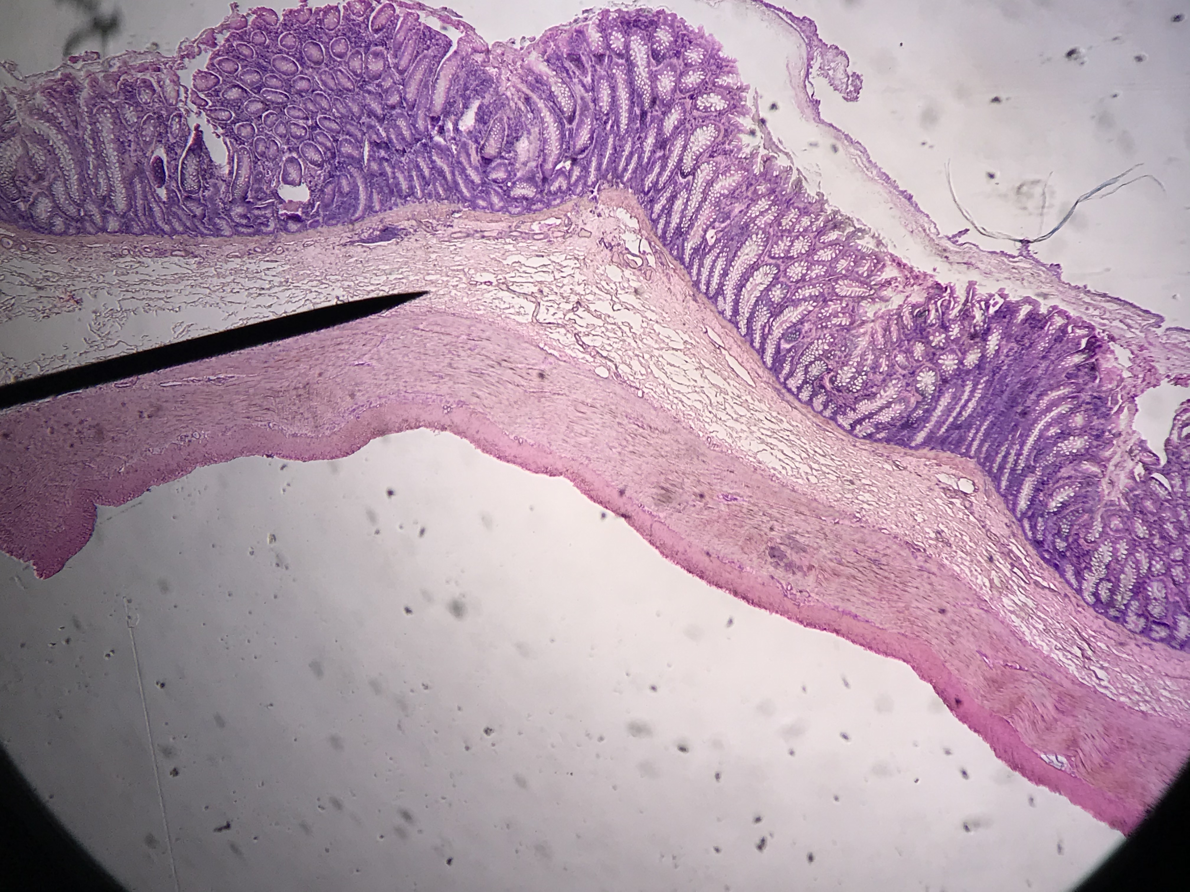
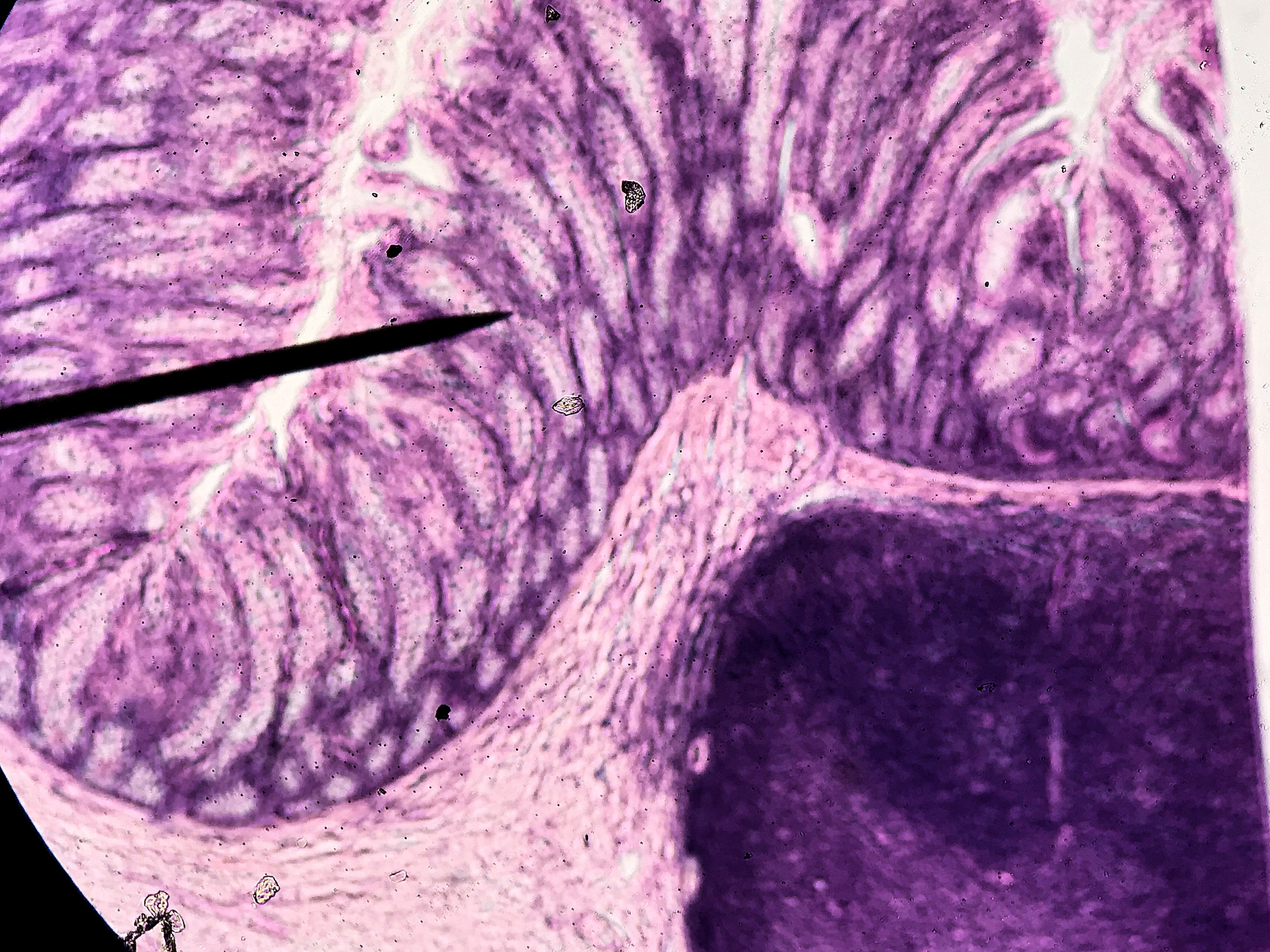
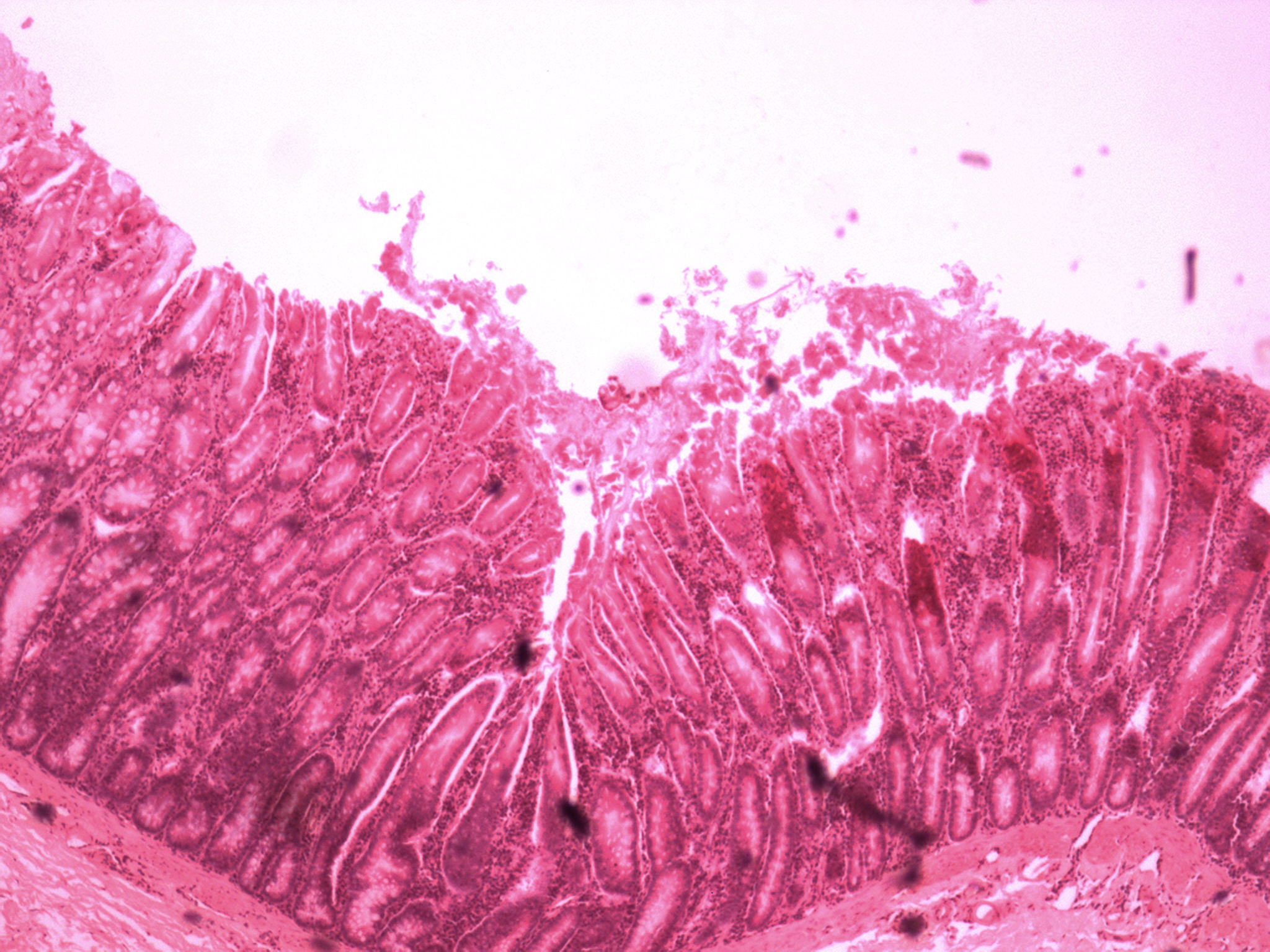
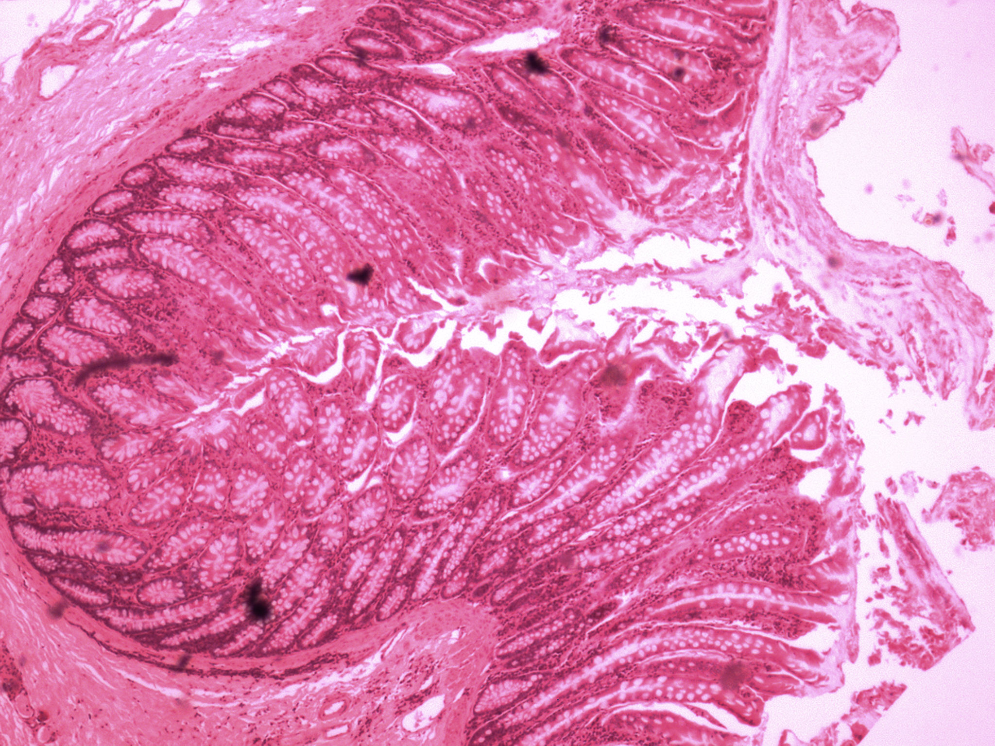
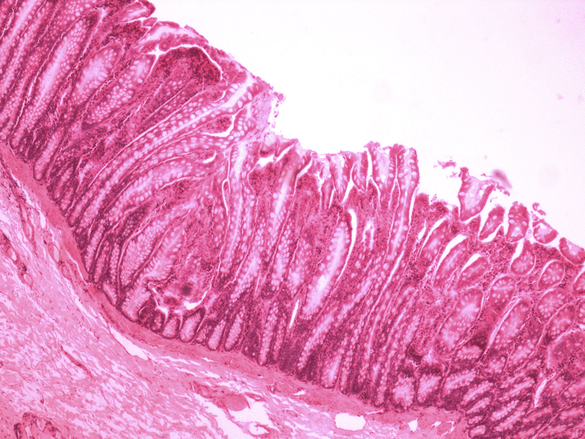
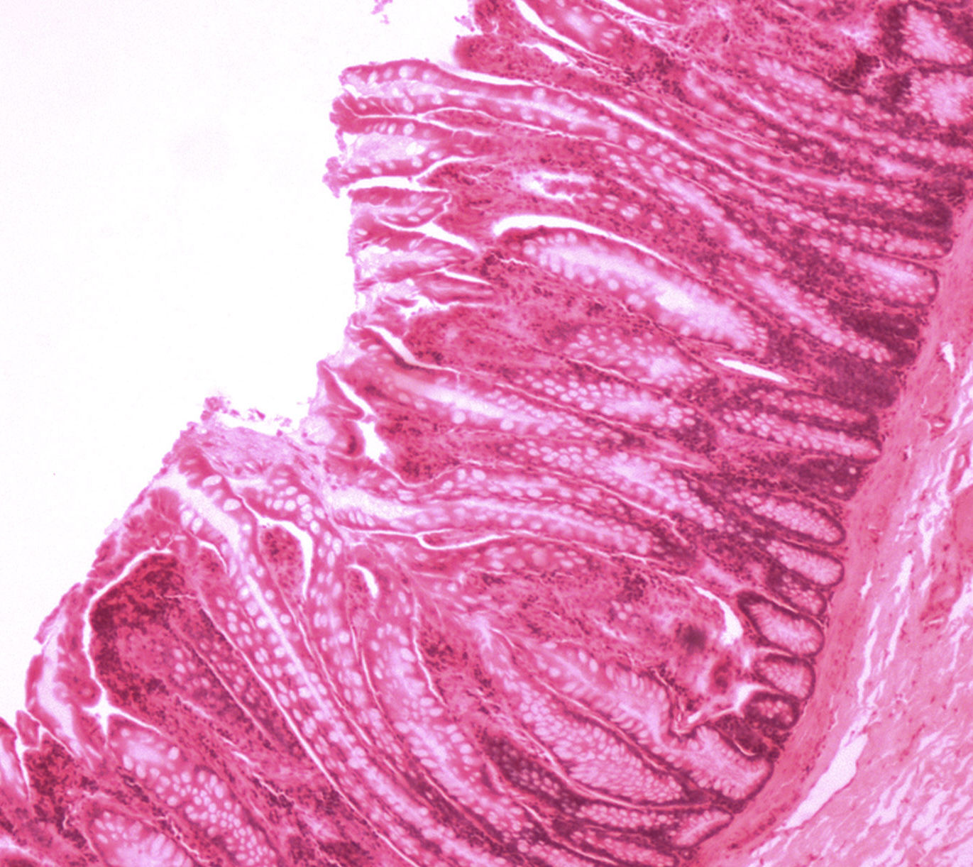
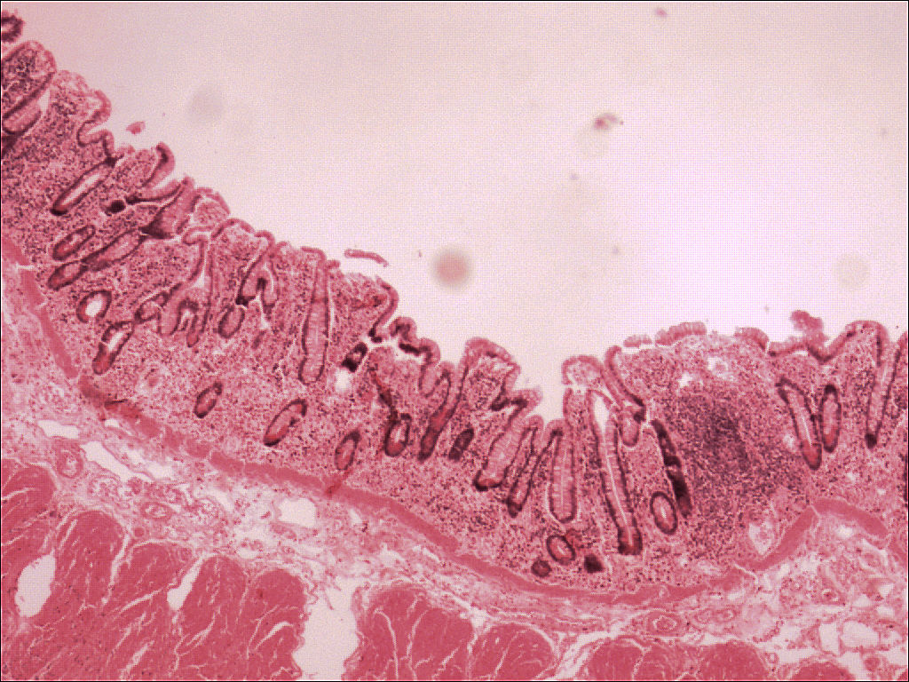
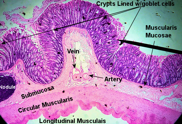
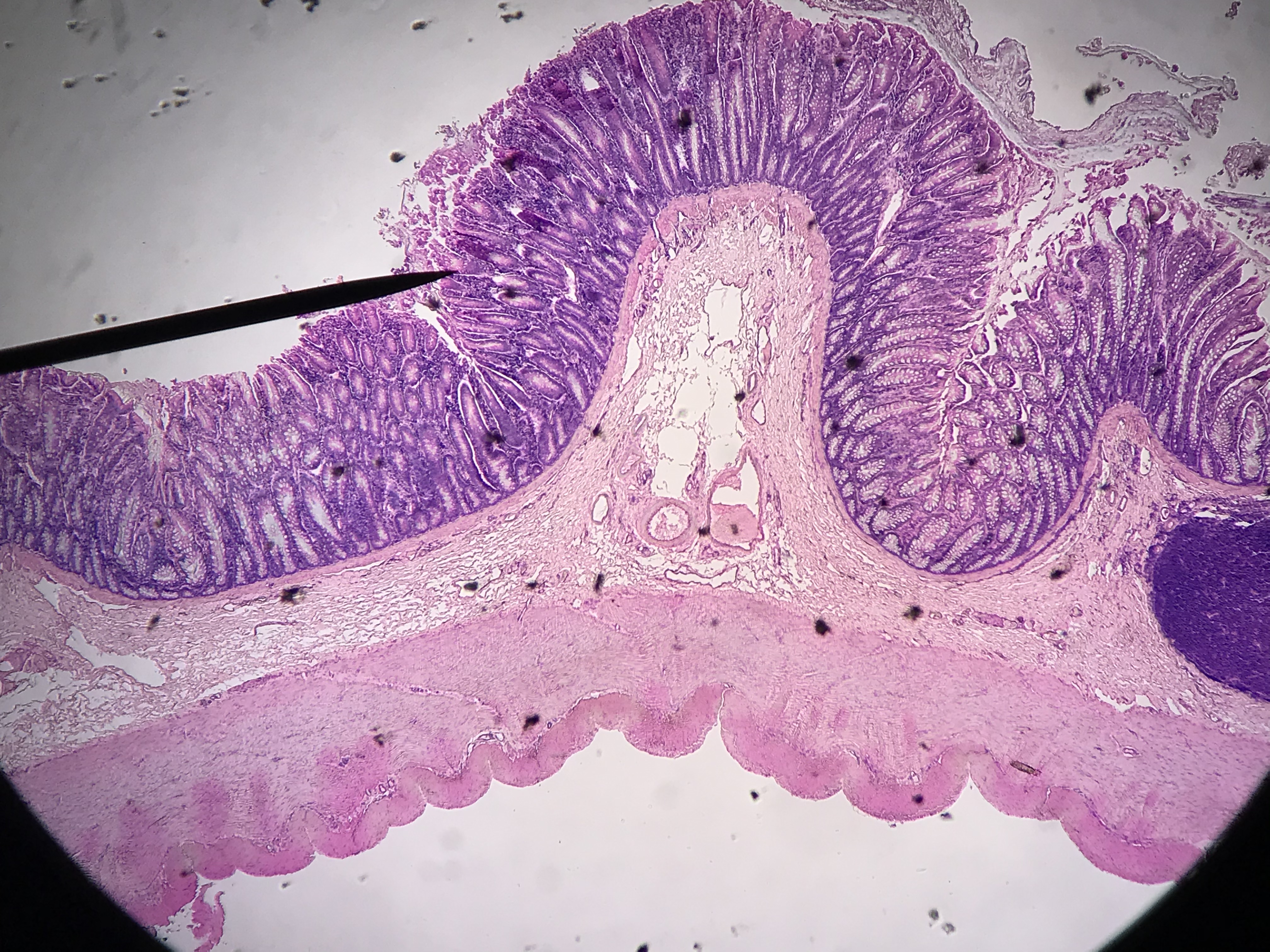
The colon is interesting to compare to the other 3 sections of the GI tract. The muscularis has deep crypts that are lined with goblet cells. The goblet cells produce a lot of mucus which help lubricate/clump the fecal matter and stem cells in the crypts replace cells lost due to friction from the mucosal layer. Lymph nodules can be observed in the submucosa but it is nothing like the ileum or appendix. The muscularis sits below the submucosa. However, the longitudinal layer may be missing depending on where the slide was taken from. The The first 9 slides were taken by me in the spring of 2021 using my phone. The rest were taken by me in 2009. The microscope camera had a red filter on it; therefore, the slides are a bit on the magenta side. Scroll through the slides and compare the slides to the labeled slide. See if you can find the slide with a lymph nodule. This model shows what the layers should be also.
Labeled Image Student Images
1 / 14
Colon
scanning
2 / 14
Colon
scanningr
3 / 14
Colon
scanning
4 / 14
Colon
scanning
5 / 14
Colon
High Power
6 / 14
Colon
Low Power
7 / 14
Colon
Low Power
8 / 14
Colon
Low Power
9 / 14
Colon
Low Power
10 / 14
Colon
High Power
11 / 14
Colon
High Power
12 / 14
Colon
Low Power
13 / 14
Colon
High
14 / 14
Colon
Scan