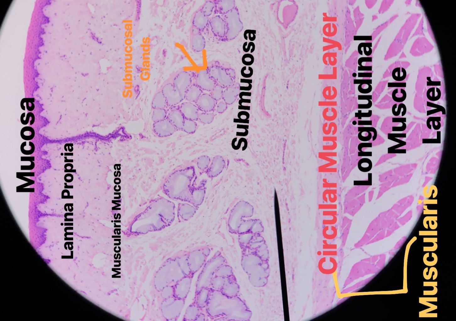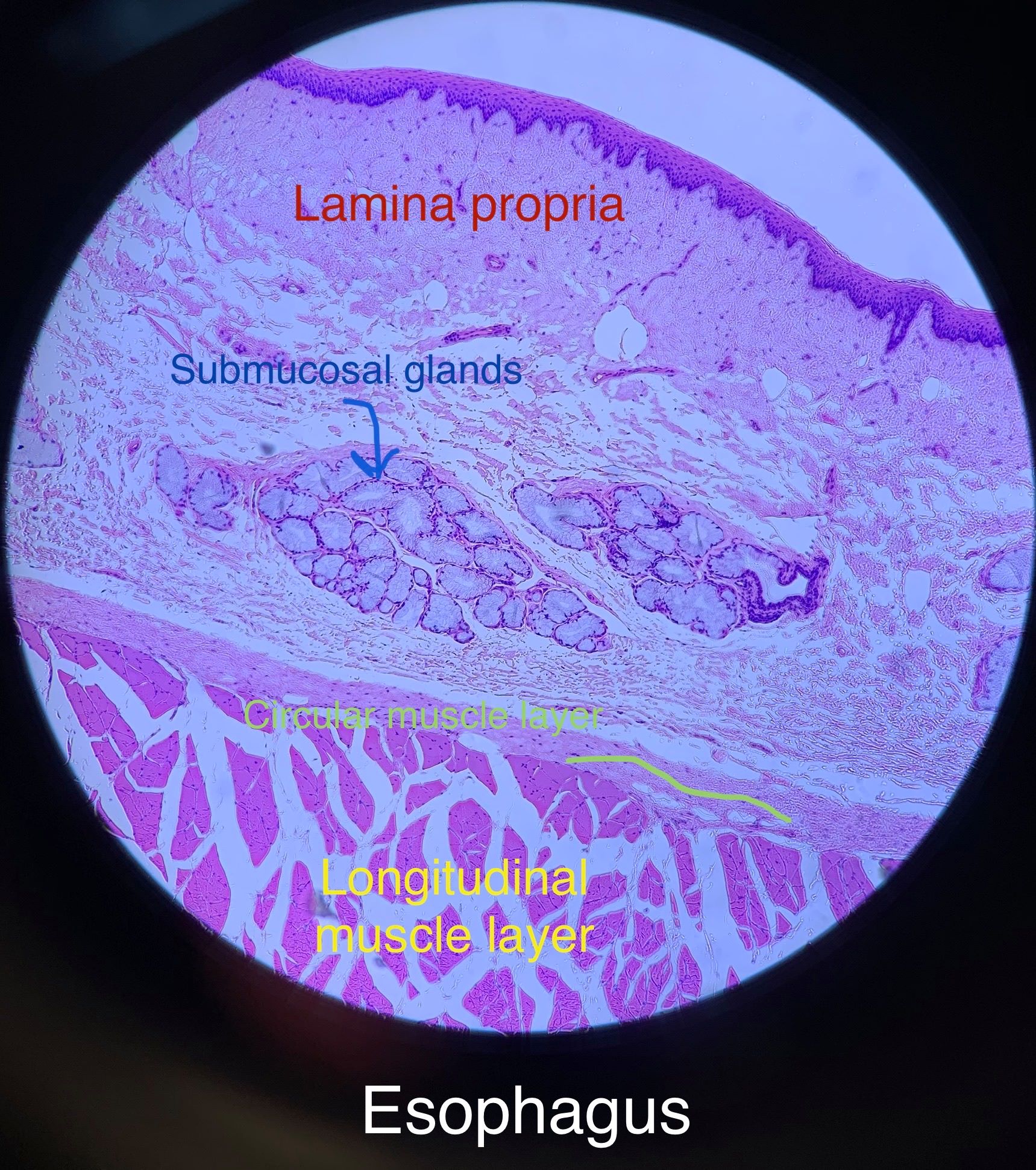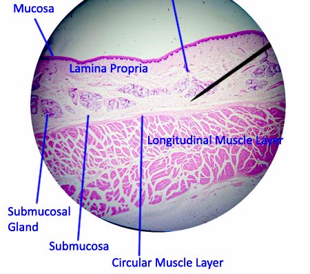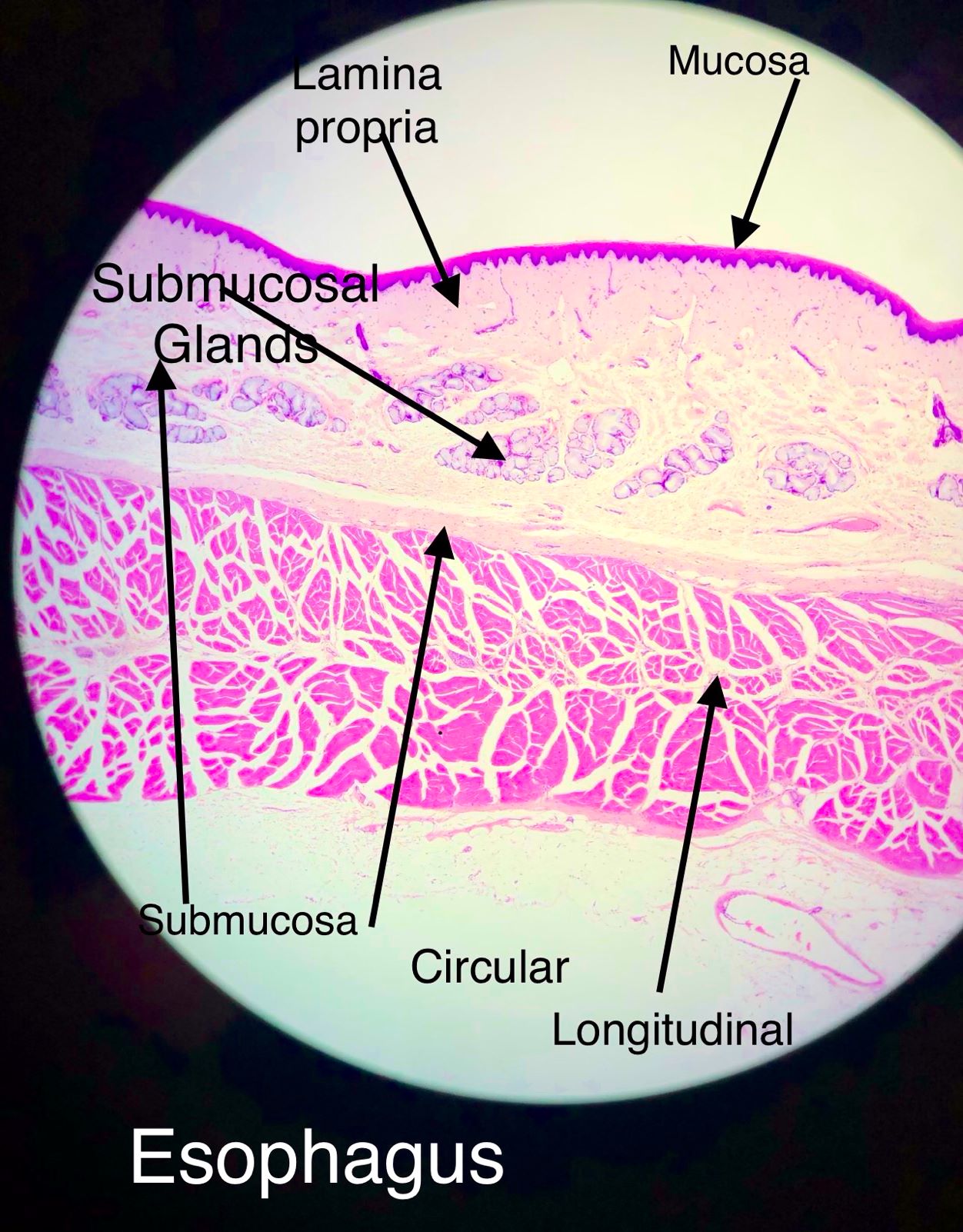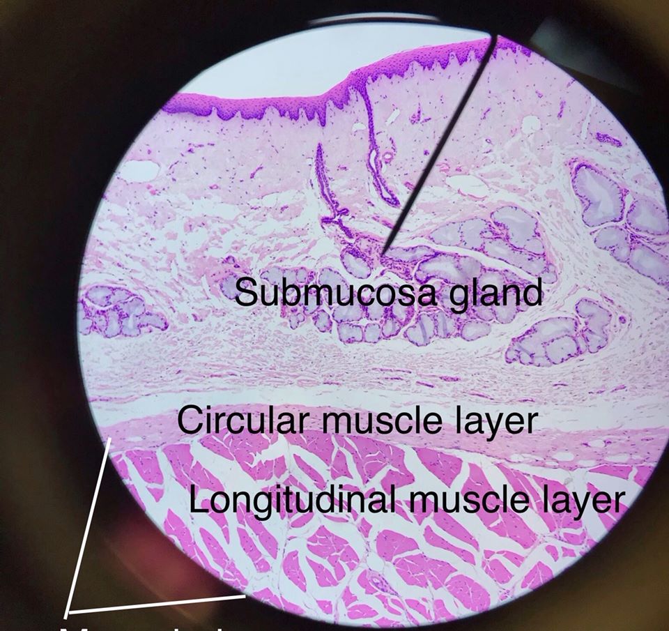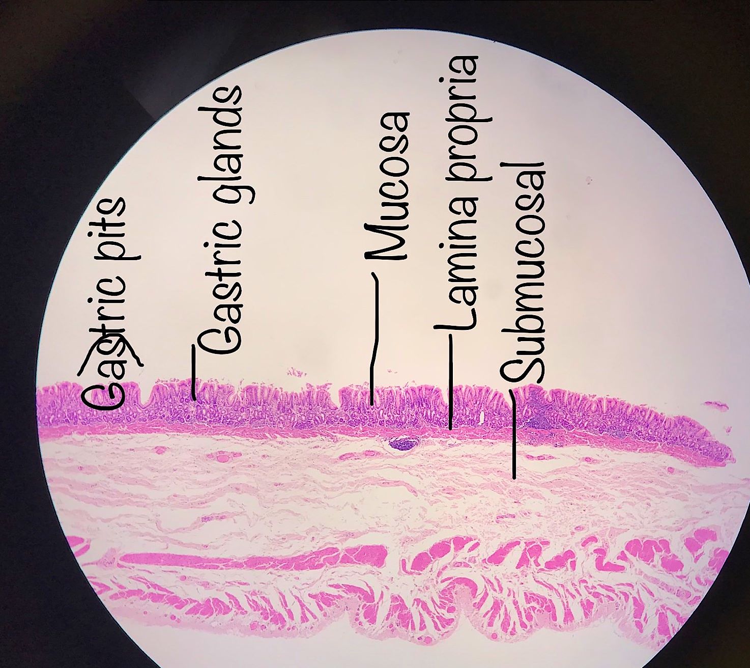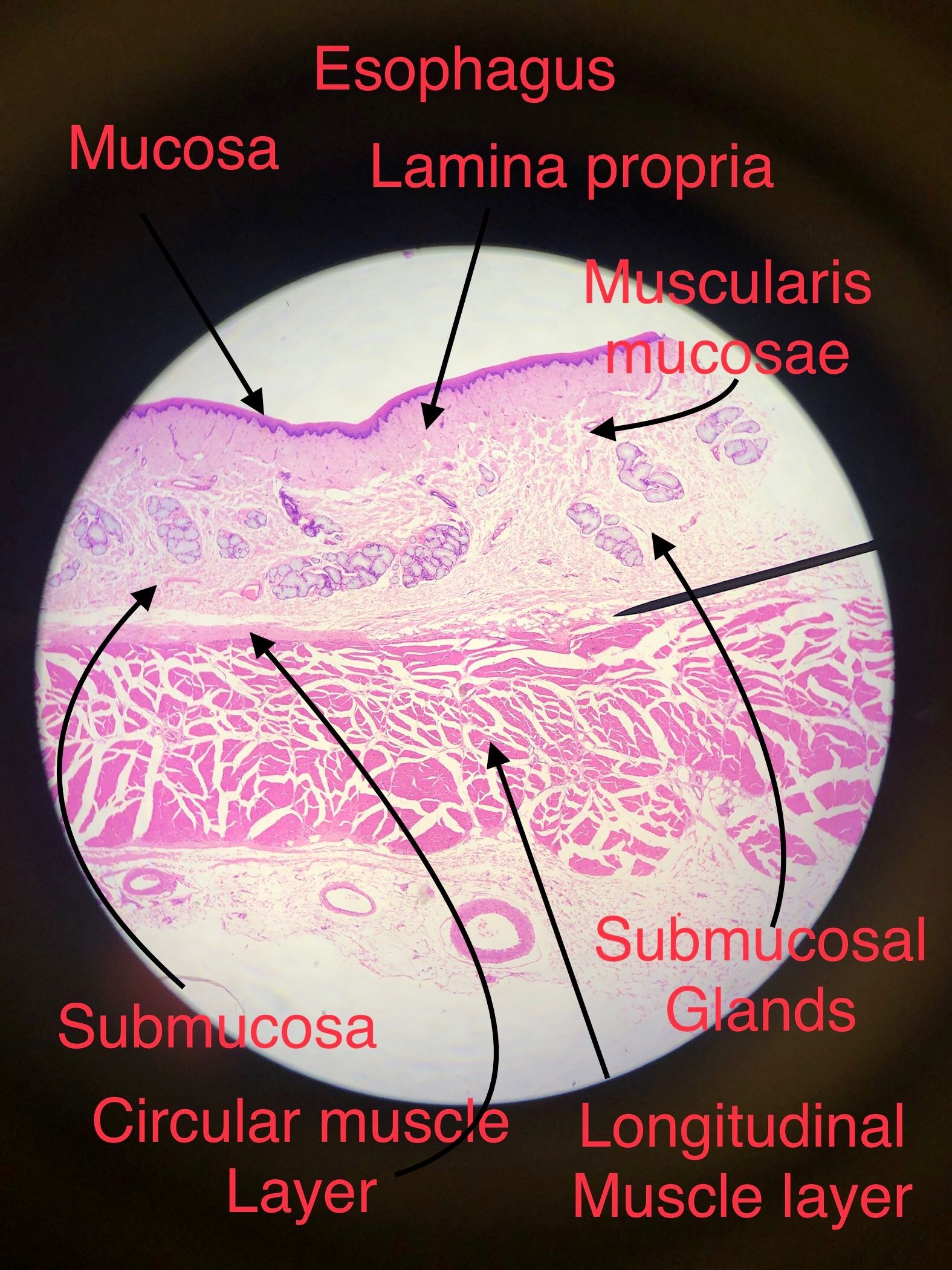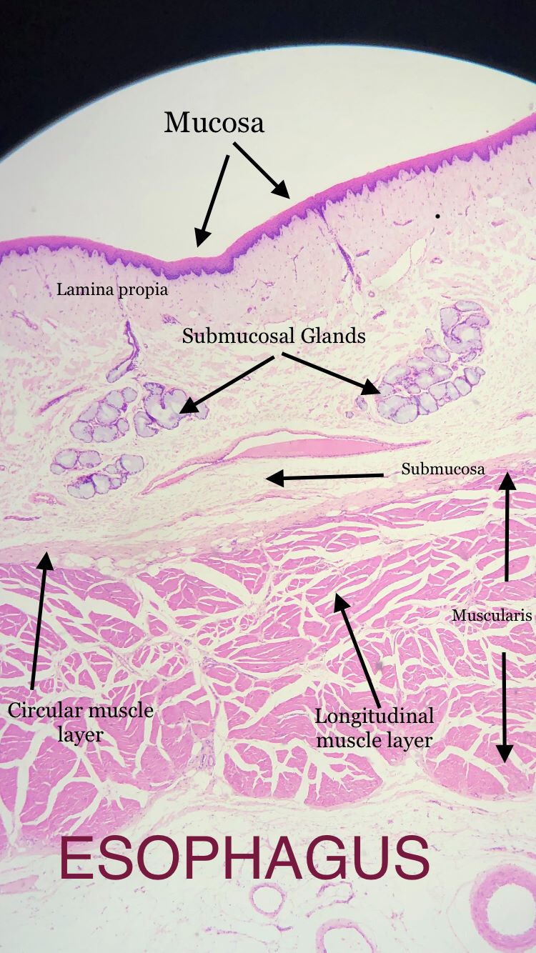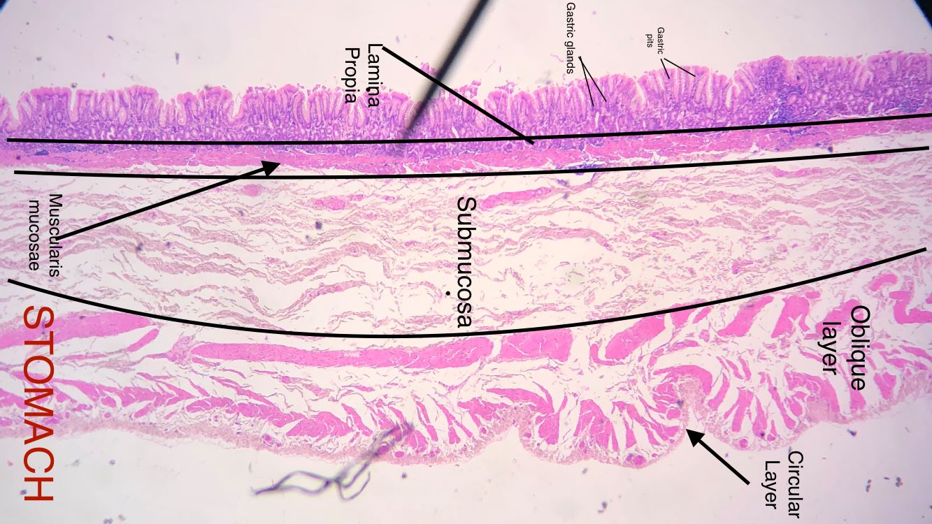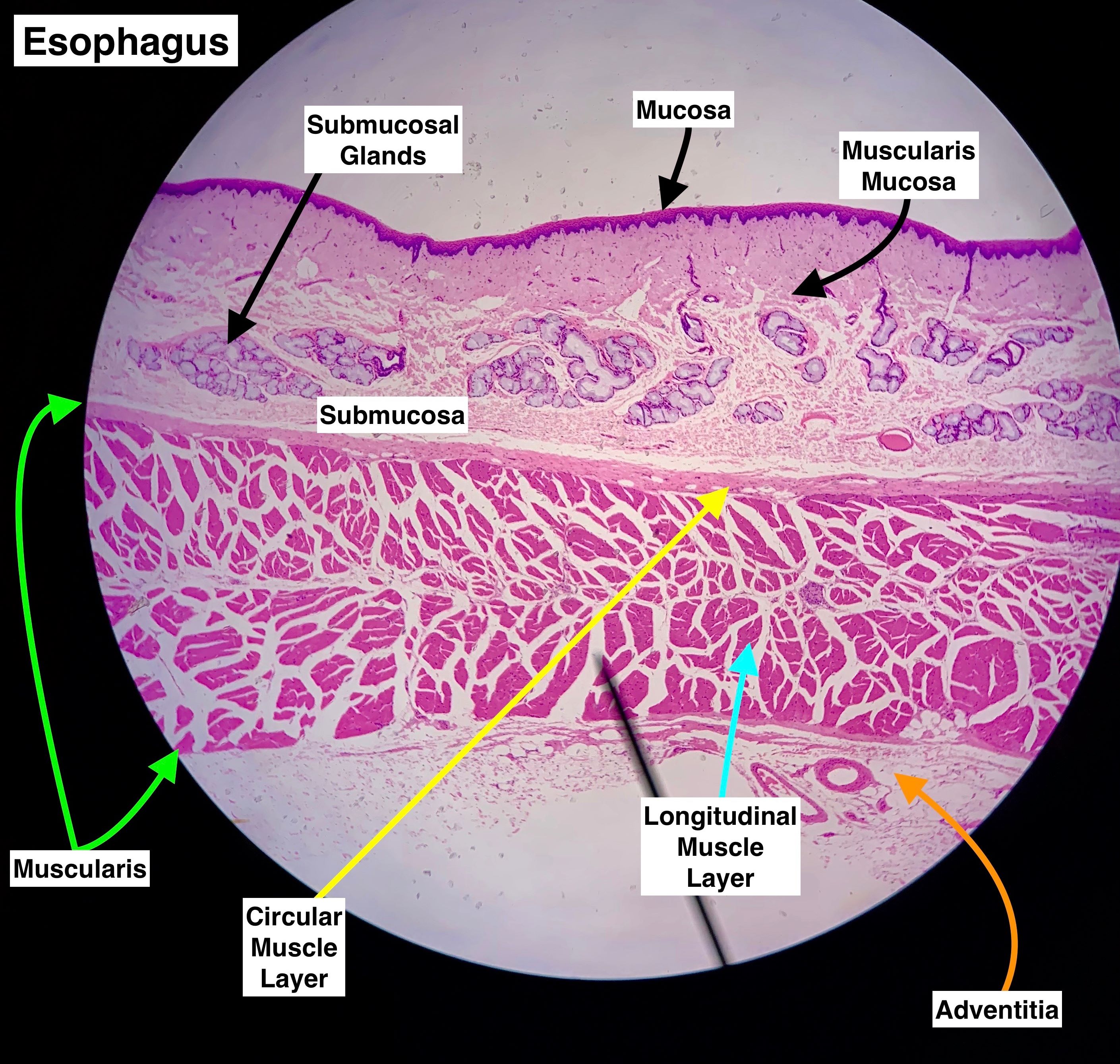
The lumen of the esophagus is surrounded by stratified squamosal epithelial tissue and this makes up the mucosal layer epithelial tissue interspersed with esophageal glands that secrete a protective mucus. Underneath that is the lamina propria which anchors the basement membrane with the submucosa. In this layer the muscularis mucosae is found and this layer of muscle is continuous through the whole tract. The next layer that we will discuss is the submucosal layer which is made of dense irregular connective tissue. This layer contains nerves, lymph vessels and blood vessels which supply the tissue with oxygen and take absorbed nutrients away. It also has very prominent submucosal glands which as the name implies also secrete mucus. The next layer is the muscularis and this contains typically 2 bands of muscles; an inner circular layer and an outer longitudinal layer. These layers propel the food with rhythmic contractions called peristaltic contractions. The outer layer of the esophagus is the adventitia which is made of dense irregular connective tissue. This is different from the rest of the digestive system; those organs have a serosal membrane around them so their outer layer will be a serosa.
The pictures on this page were taken in the spring of 2018 and fall of 2019. Scroll through the images comparing them to the static image. Select one and draw it. Also see if you can find the one where they switched the layers of the muscularis. This model shows what the layers should be also.
| Lab Book Image | Student Images |
|---|---|
|
|
|