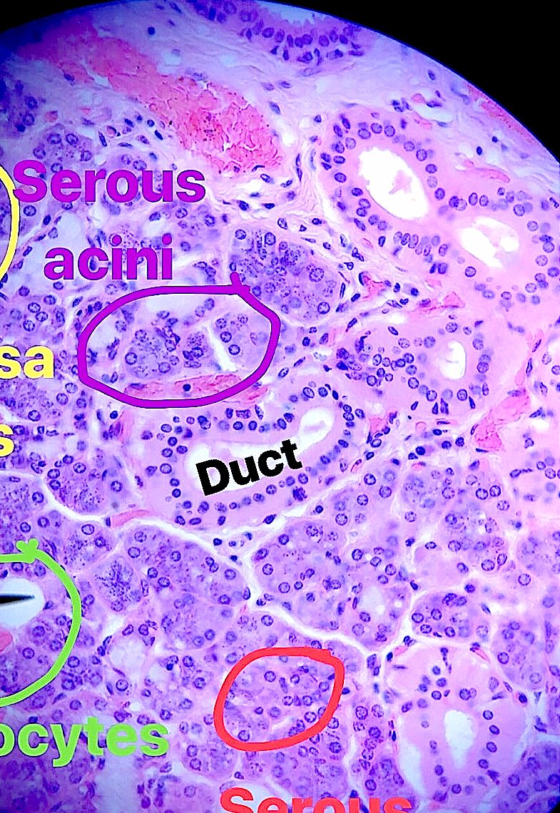
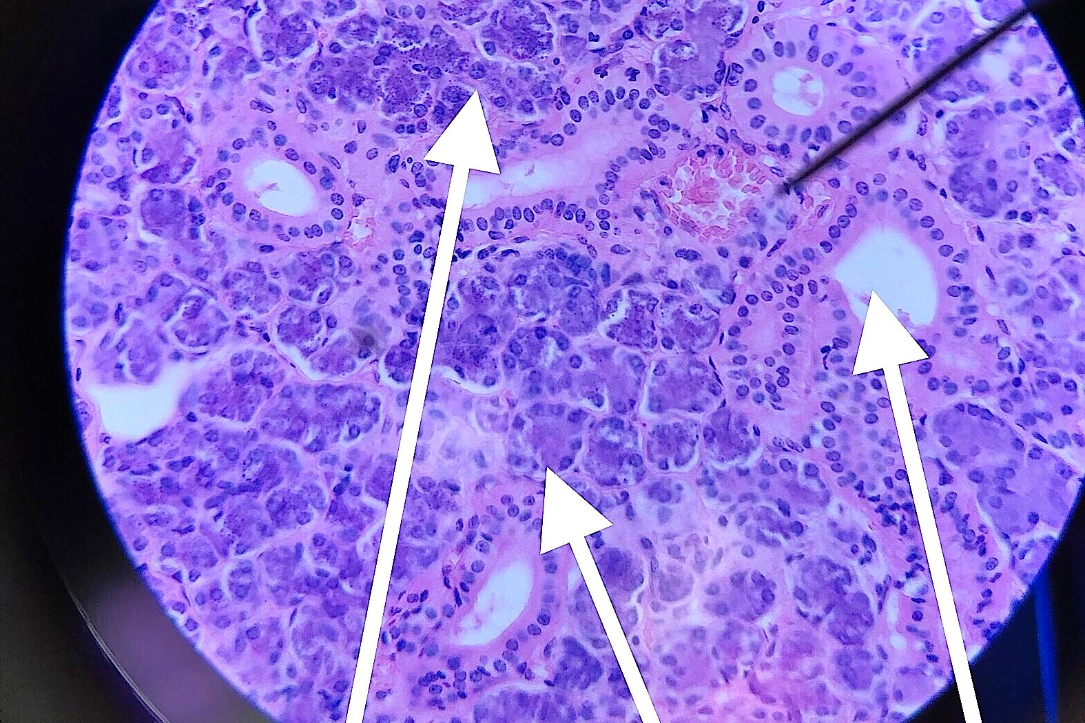
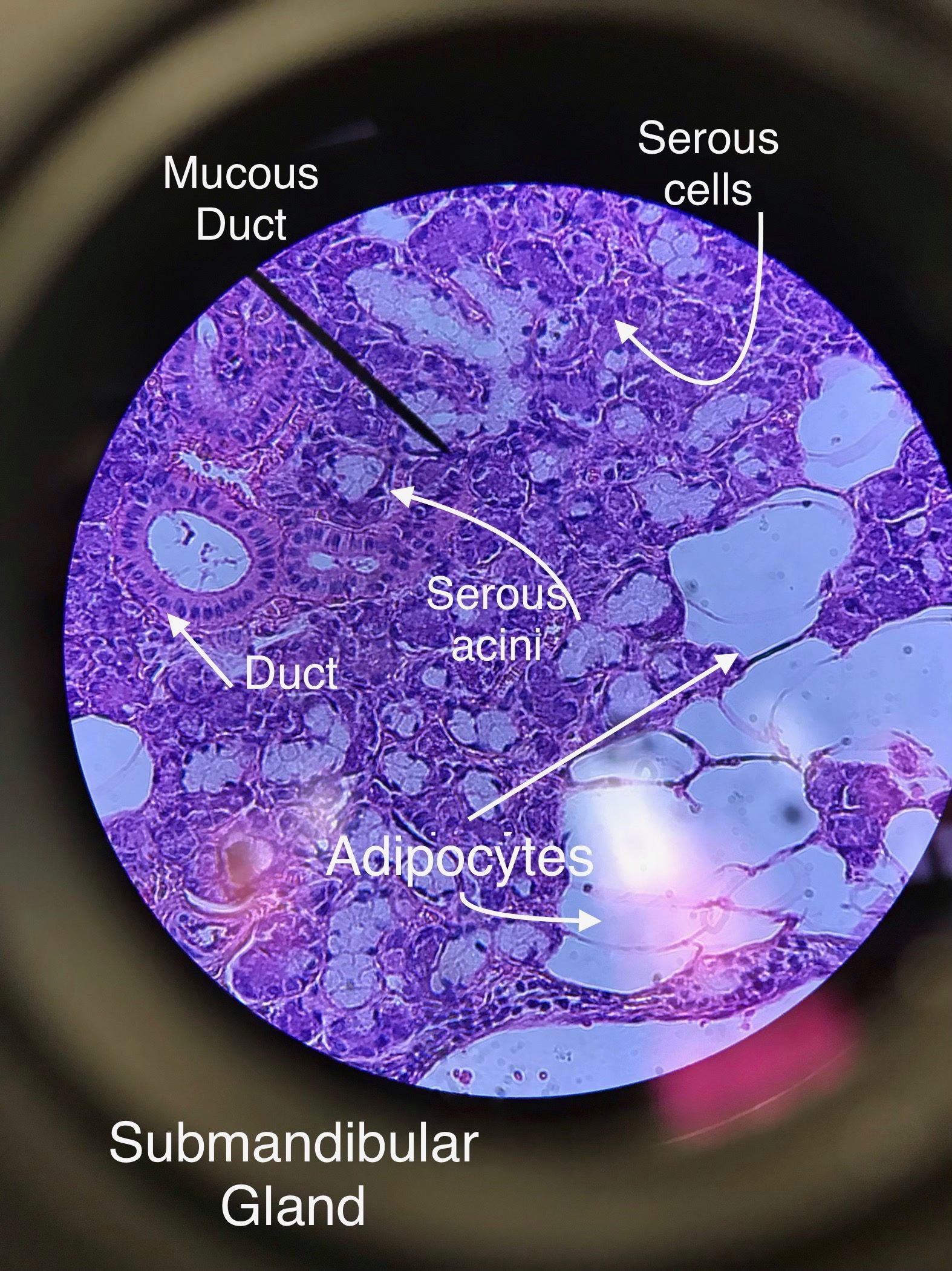
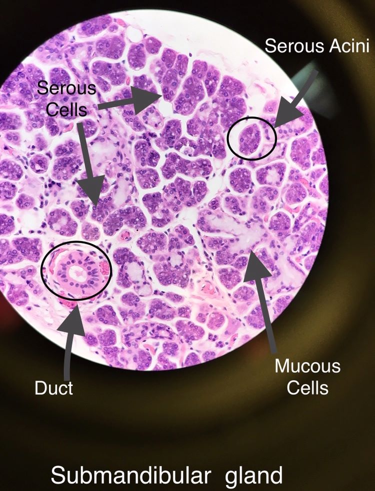
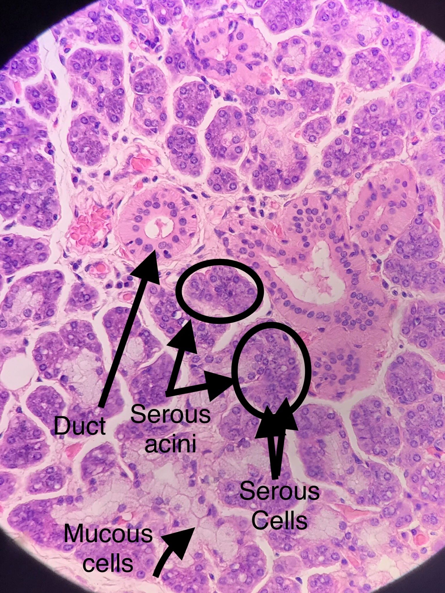
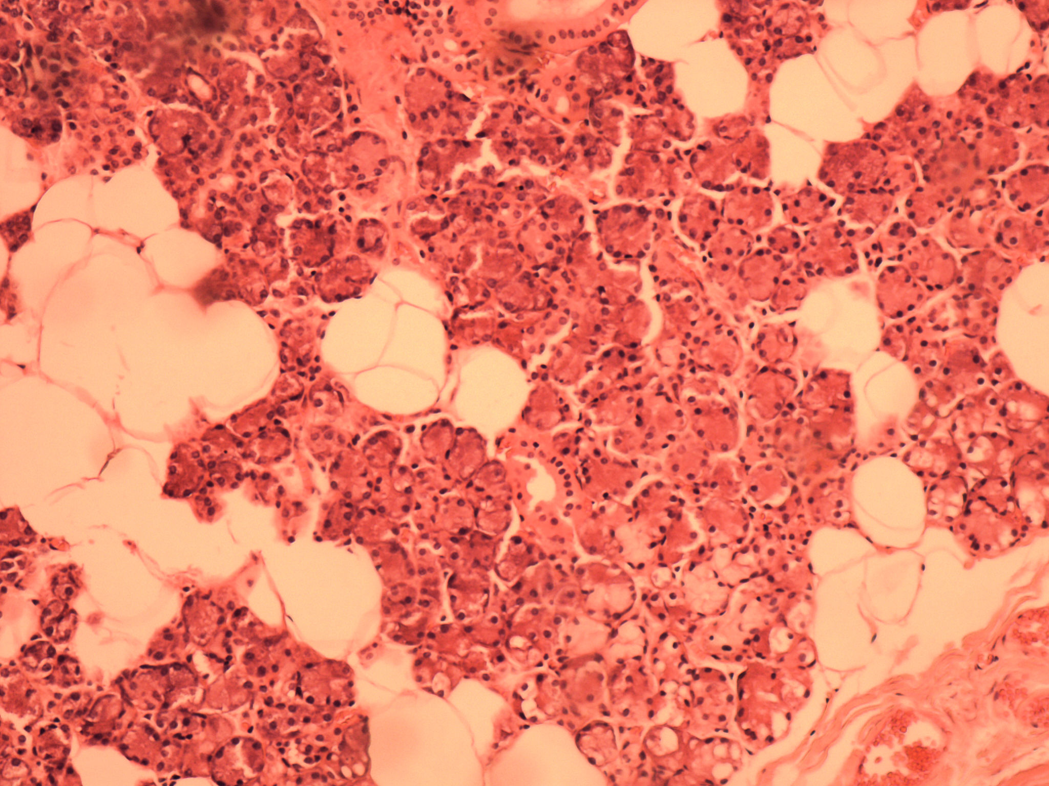
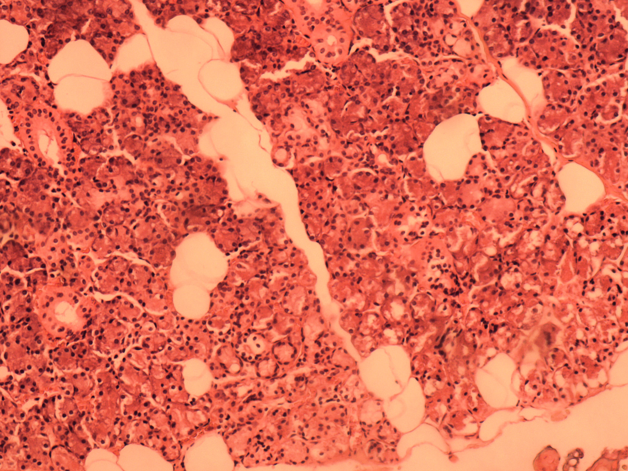
There are two types cells that are visible in the salivary glands. Serous cells produce salivary amylase and lysozyme, an enzyme that damages bacterial walls. Serous cells are arranged in clusters called acini. Mucous cells are the other cell present and as the name implies, these cells make mucus that helps to lubricate the food. These cells tend to be larger and white in color. Secretions from both cell type drain into salivary ducts. These are made up of cuboidal epithelium (both simple and stratified). The parotid gland is made up predominantly of serous cells whereas the sublingual gland is predominantly mucous cells. The submandibular gland is divided into units called adenomers. These adenomers will contain either mucous cells or serous cells. These pictures were taken by bio 139 students in the spring of 2018 and fall of 2019. Scroll through the pictures and compare them with the labeled picture. Select one and draw it.
| Lab Book Image | Student Images |
|---|---|
|
|
|