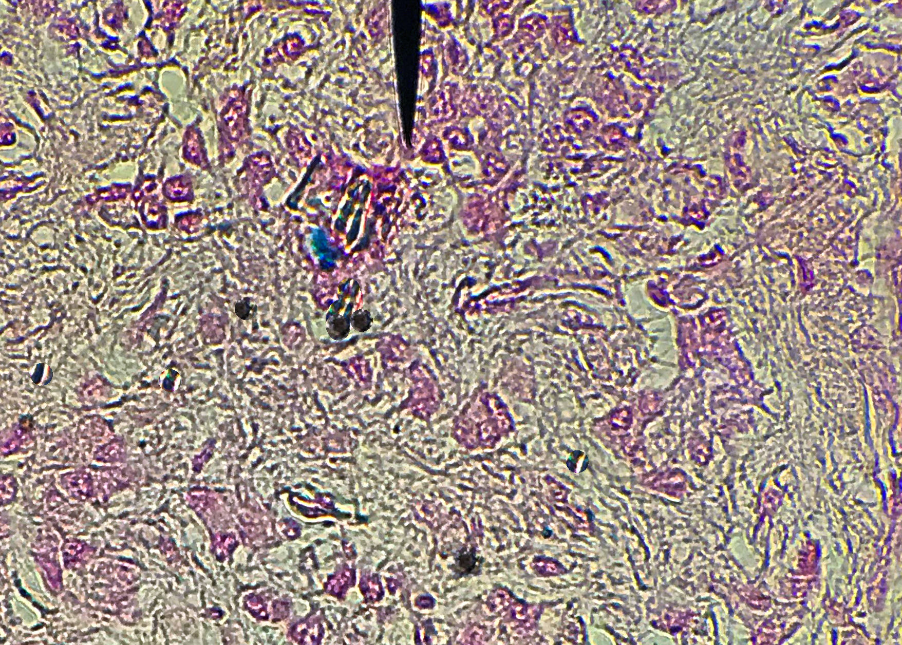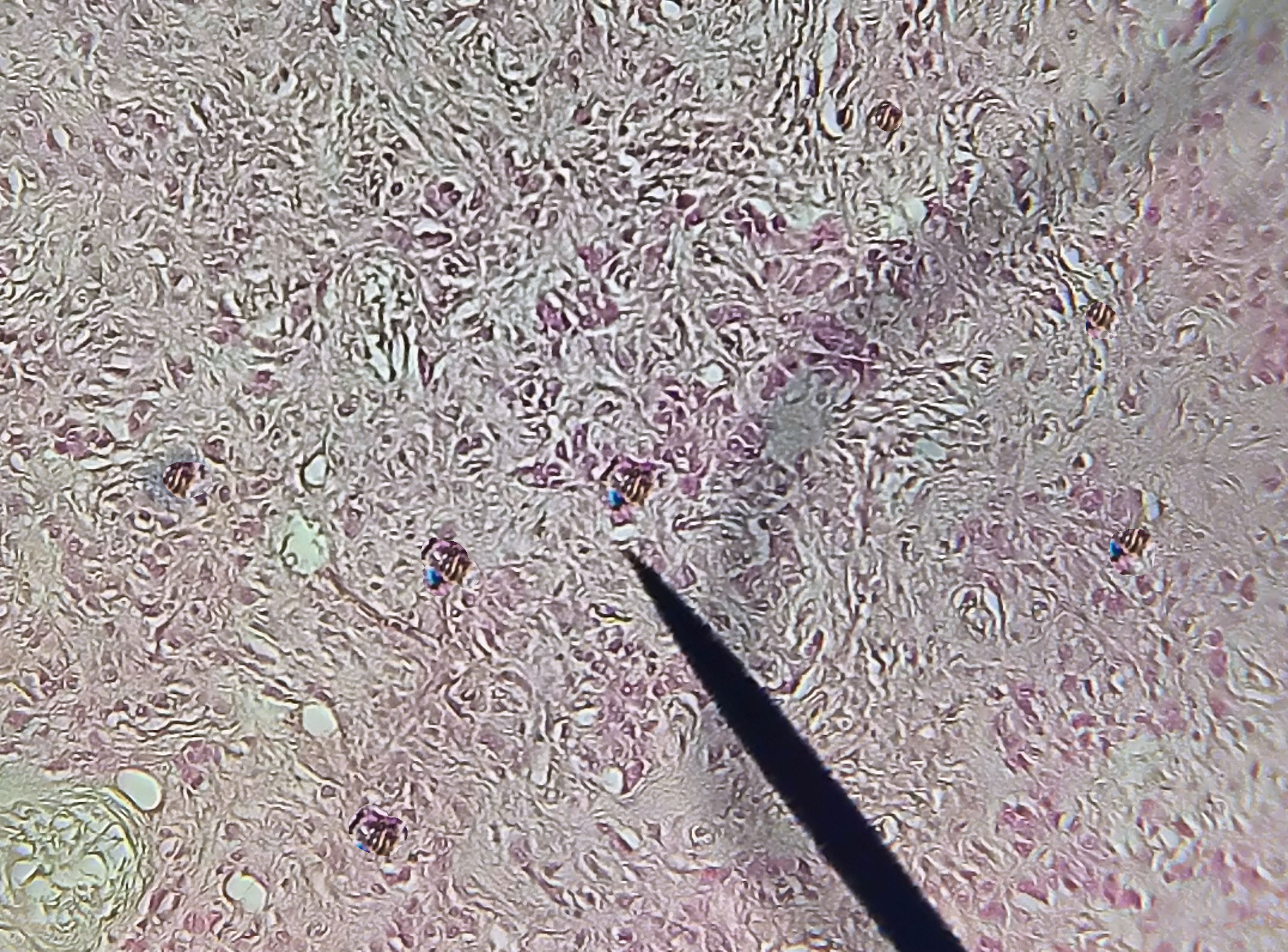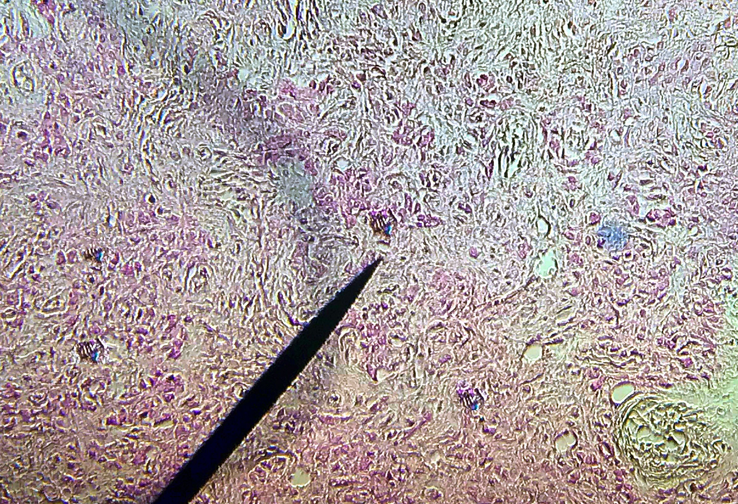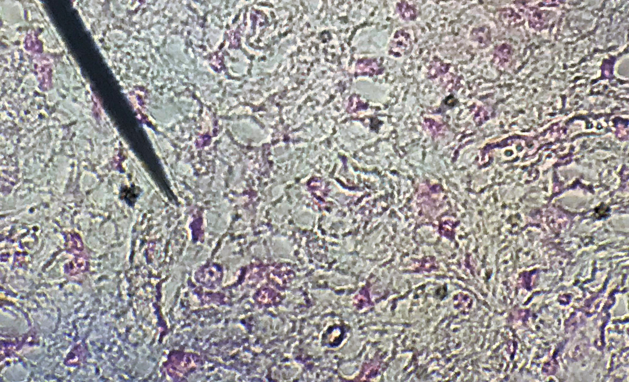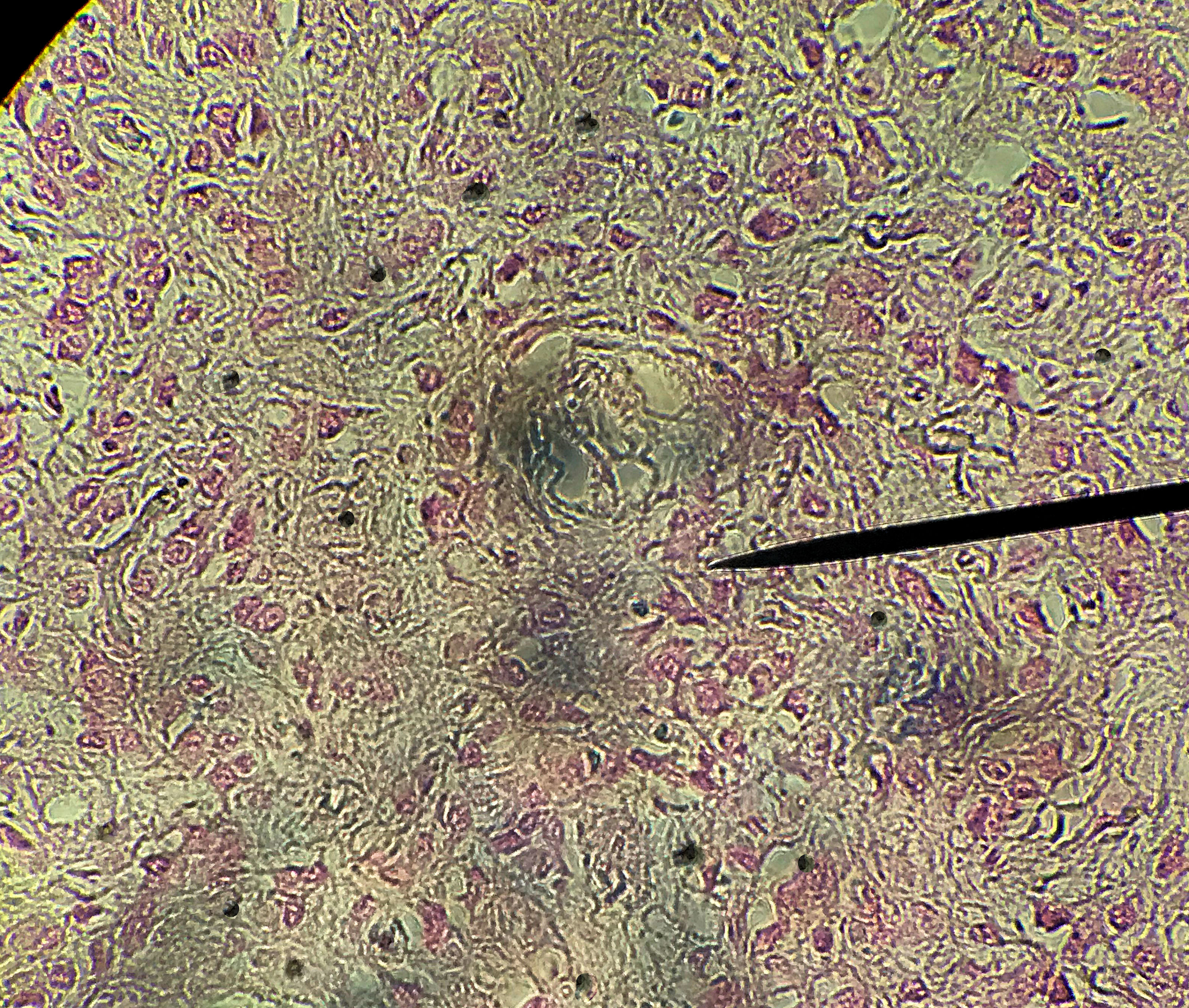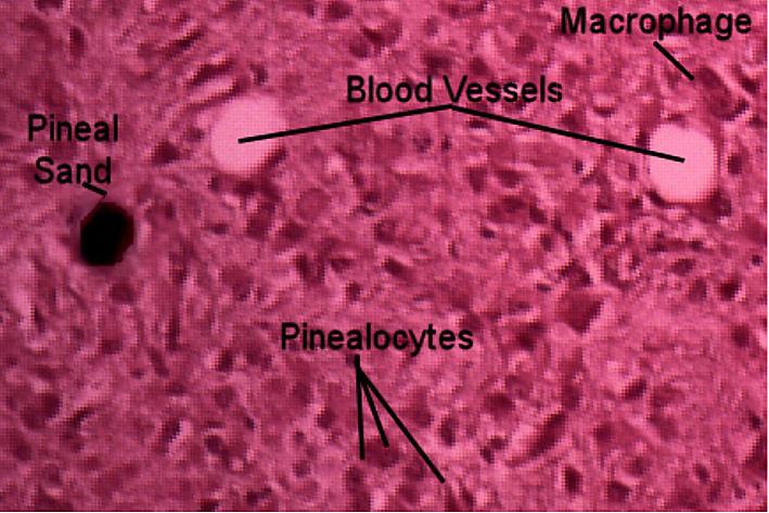
The pineal gland is a pine nut size gland that is found in the epithalamus and its function is to help establish circadian rhythms through the secretion of melatonin which is secreted in the dark. Melatonin's main function in humans is to promote sleepiness and to get your body ready for sleep. The pineal gland is made up mostly of pinealocytes which produce melatonin. The pinealocytes are surrounded by astrocytes which aid in the environmental regulation of the pineal gland. Since the pineal gland is outside the brain blood barrier, it is also possible to see blood vessels on a histological slide. As we age, the pineal gland calcifies. These calcium deposits are referred to as pineal sand. This page has endocrine histology of the pineal under high power. Slides which were taken by bio 139 students from spring of 2018 to fall of 2019 are in the right hand side and your lab book's picture is on the left hand side.
Compare those pictures to the lab book pictures by scrolling trough the student pictures using the yellow arrows. Then draw the histology as instructed by your teacher.
| Lab Book Image | Student Images |
|---|---|
|
|
Pineal Gland high Power |