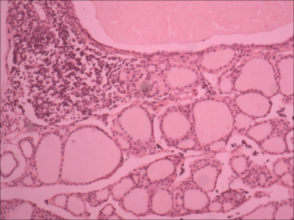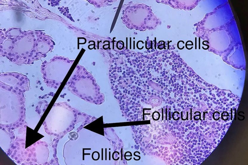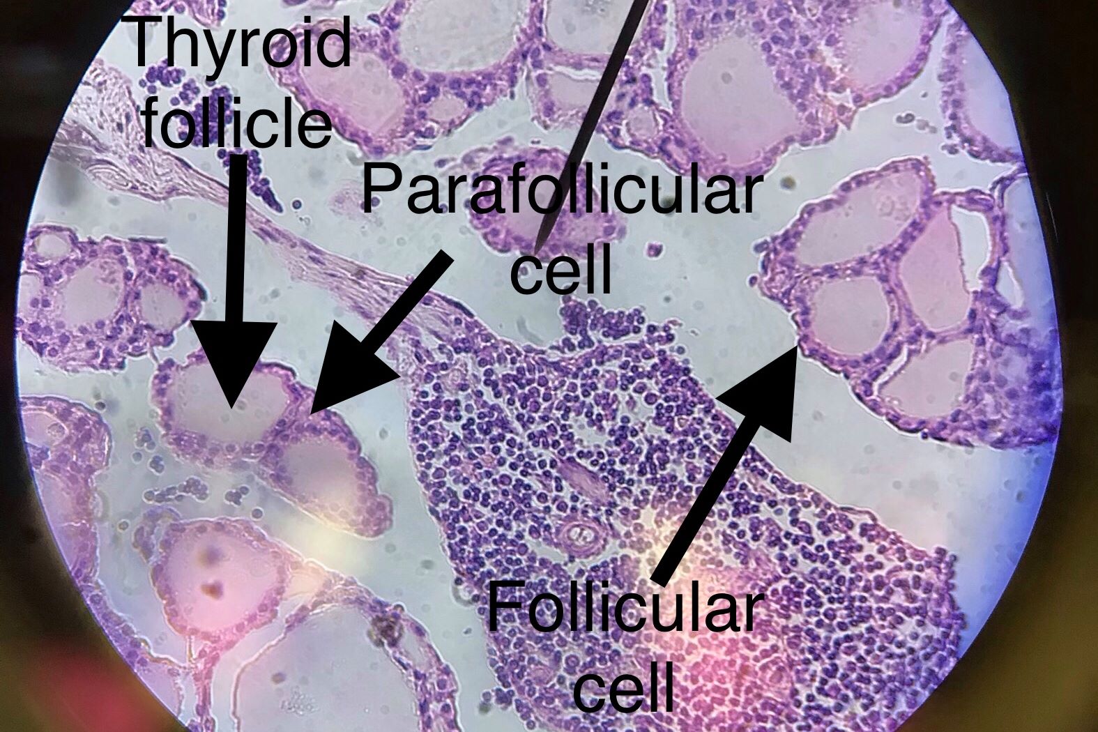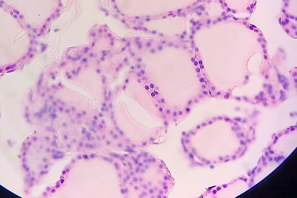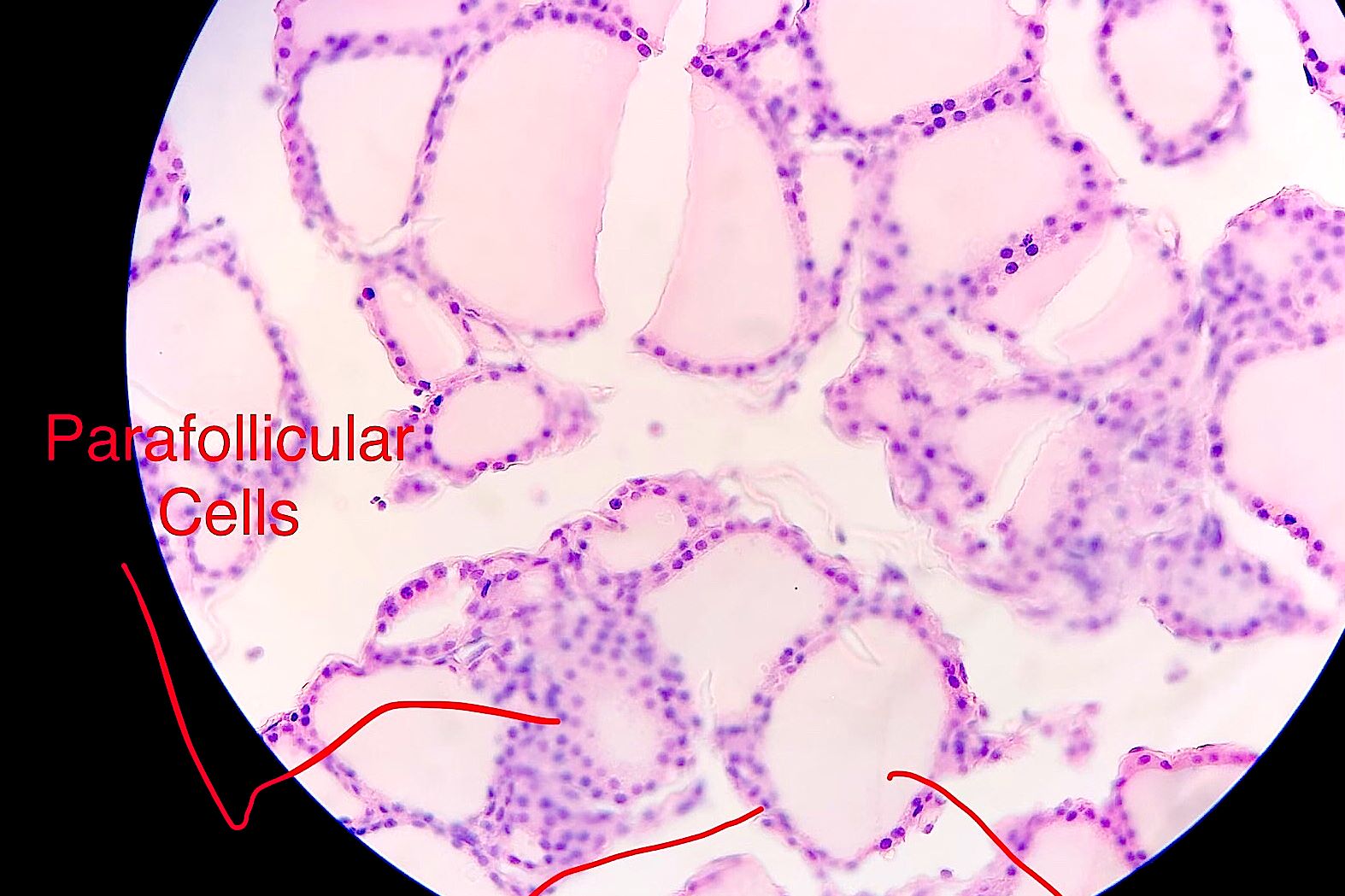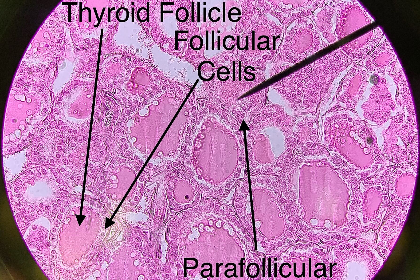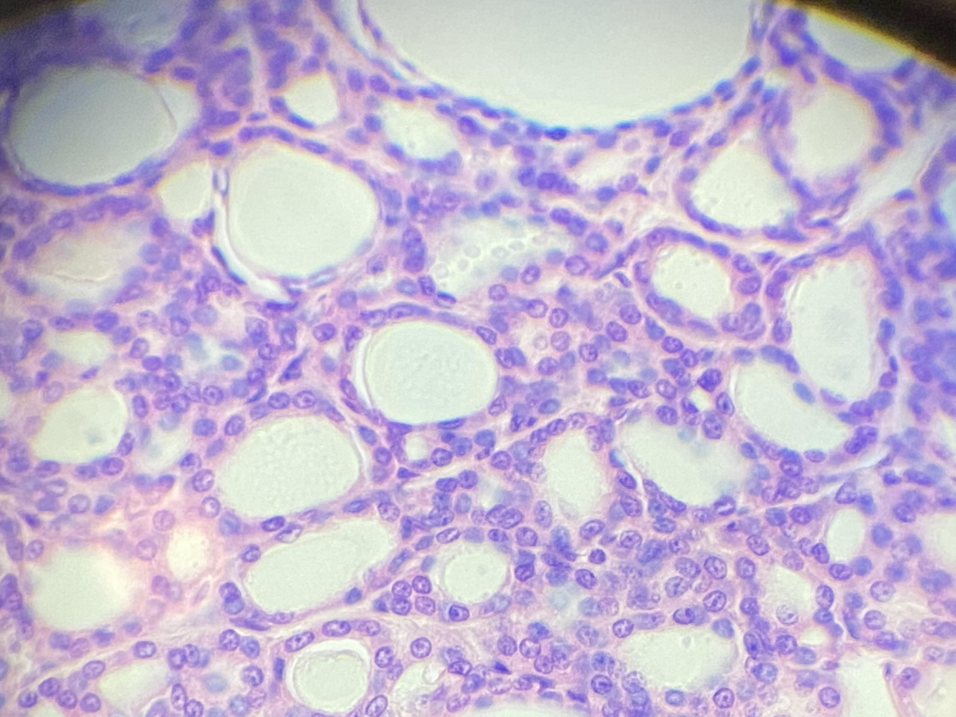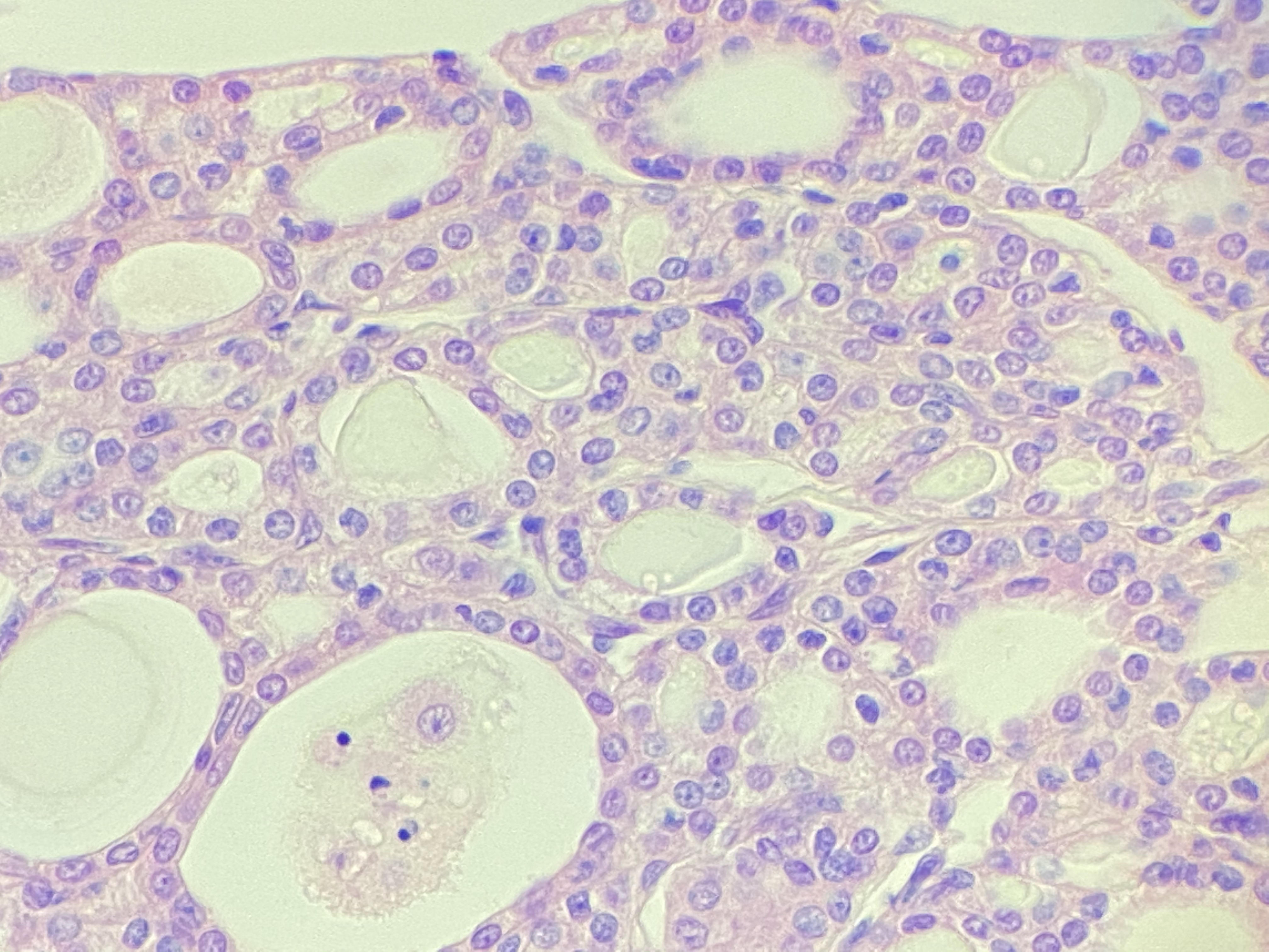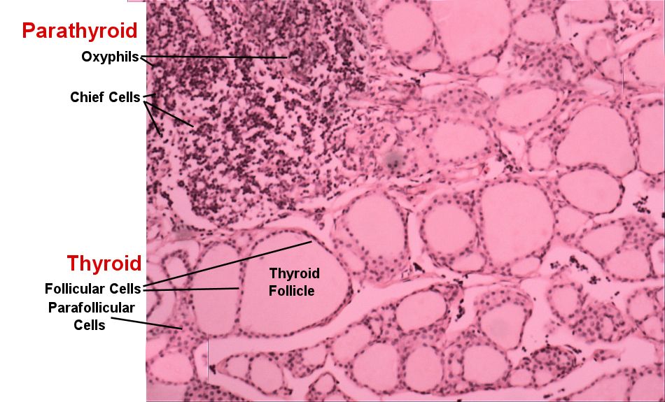
This page has endocrine histology of the thyroid gland under low power. The most prominent structure of the thyroid gland are the thyroid follicles which is where thyroid hormone, a metabolic hormone, is assembled and stored. The thyroid follicles are surrounded by the follicular cells which produce thyroid hormone. Between the thyroid follicles you will observe the parafollicular cells. These cells produce calcitonin which is a hormone that increases the activity of osteoblasts in your bone, lowering blood calcium and increasing bone density. The parathyroid glands are located on the posterior surface of the thyroid gland and are often observed with the thyroid gland. There are two sets of cells in the parathyroid glands. Chief cells are small cells that stain dark purple with lots of granules. They produce parathyroid hormone which functions to maintain blood calcium above 8 mg/dl. You will also observe the larger oxyphils cells that stain pinkish and tend to have a nuclei surrounded by cytoplasm. Their function is unknown.
Slides which were taken by bio 139 students from spring of 2018 to fall of 2019 are in the right hand side and your lab book's picture is on the left hand side. Compare those pictures to the lab book pictures by scrolling trough the student pictures using the black arrows. Then draw the histology as instructed by your teacher.
| Lab Book Image | Student Images |
|---|---|
|
|
Thyroid Gland Low Power |