1 /8
Scanning
2 / 8
Low Power
3 / 8
High Power
4 / 8
High Power
5 / 8
Low Power
6 /7
Scanning
7 / 8
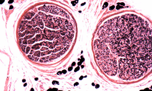
Osmic Acid, Low Power
8 / 8
Osmic Acid High Power
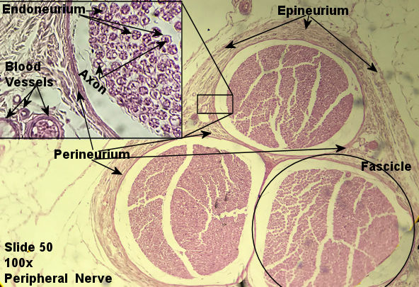
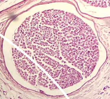
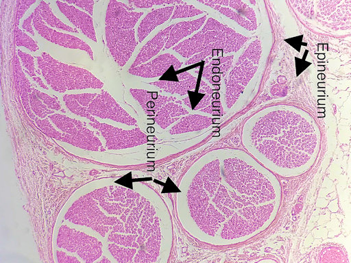
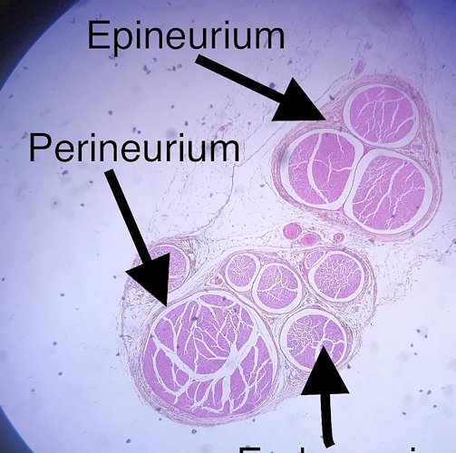
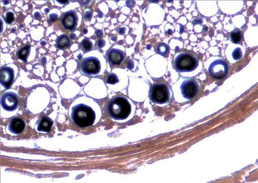
This page has the connective tissue elements of a nerve. The picture to the left is from the lab book. The picture on the right are student files from the fall of 2019.
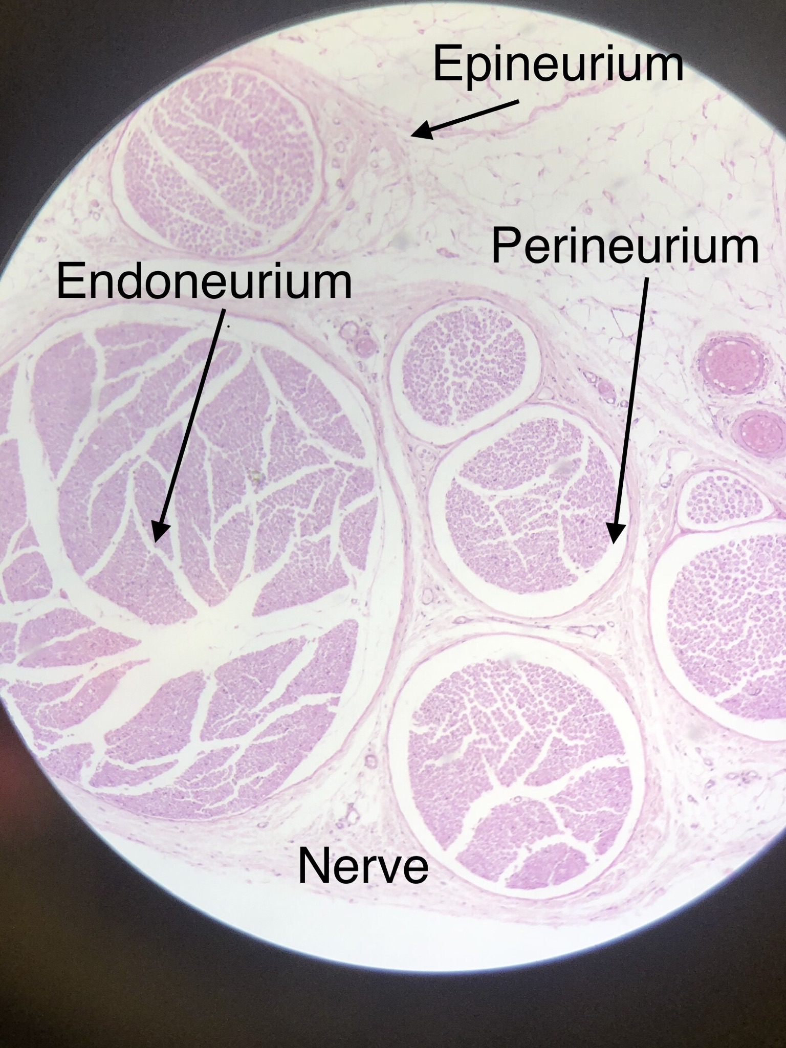
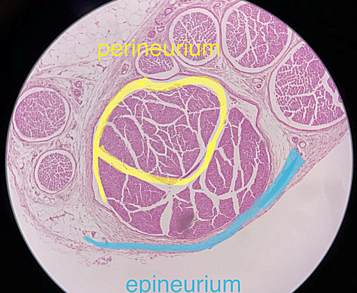
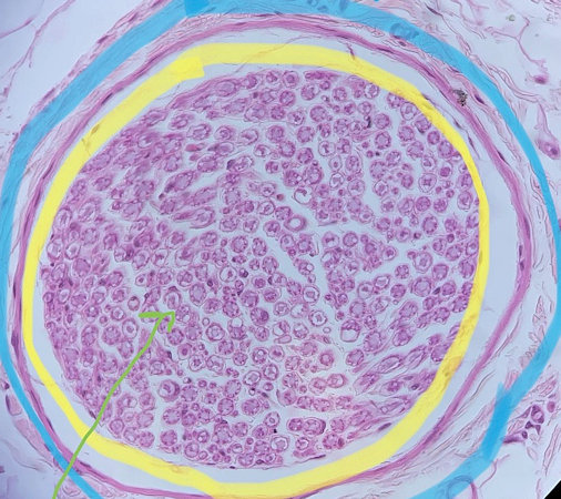
A nerve is really a collection of axons that are wrapped together and insulated much like an electrical wire. The outside of a nerve is wrapped in a layer of dense regular connective tissue called the epineurium. The epineurium is a continuation of the connective tissue elements of the central nervous system and allow room for blood vessels, lymphatic vessels, lymphocytes and fibroblasts. Axons are wrapped within the epineurium in bundles called fascicles. The fascicles are surrounded by a layer of connective tissue called the perineurium. If you look at the stain done with osmic acid above, you can see how the layers of the perineurium wrap around the neurons. Individual axons are wrapped together in an insulating sheet of connective called an endoneurium. Within the endoneurium is a fluid called endoneurial fluid that bathes the axons and provides the axons with important electrolytes (sodium) and oxygen. The endoneurium also allows for capillaries to pass through close to the axons.
Scroll through the student pictures using the arrows. Try to identify connective tissue elements. Your teacher may have you either draw or screen shot and label. The first 6 slides are the H and E stain you are used to by now. The last 2 slides are stained osmic acid. I like it better because it clearly shoews endoneurium.
| Lab Book Image | Student Images |
|---|---|
|
1 /8
Scanning
2 / 8
Low Power
3 / 8
High Power
4 / 8
High Power
5 / 8
Low Power
6 /7
Scanning
7 / 8
Osmic Acid, Low Power
8 / 8
Osmic Acid High Power
|