1 /6
High Power
2 / 6
High Power
3 / 6
High Power
4 / 6
High Power
5 / 6
High Power
6 / 6

High Power
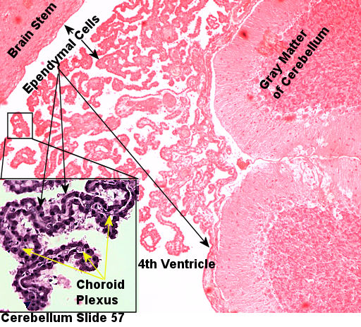
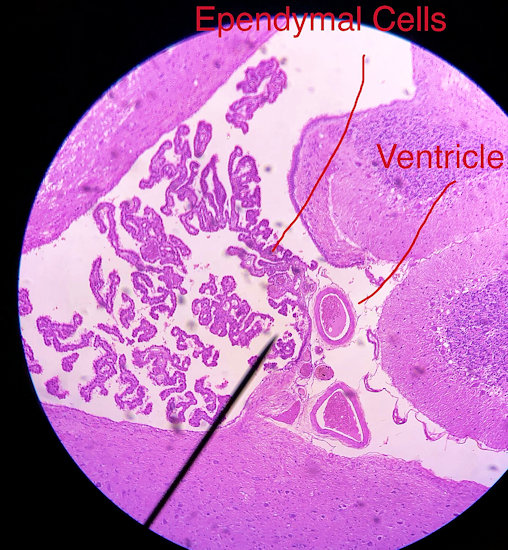
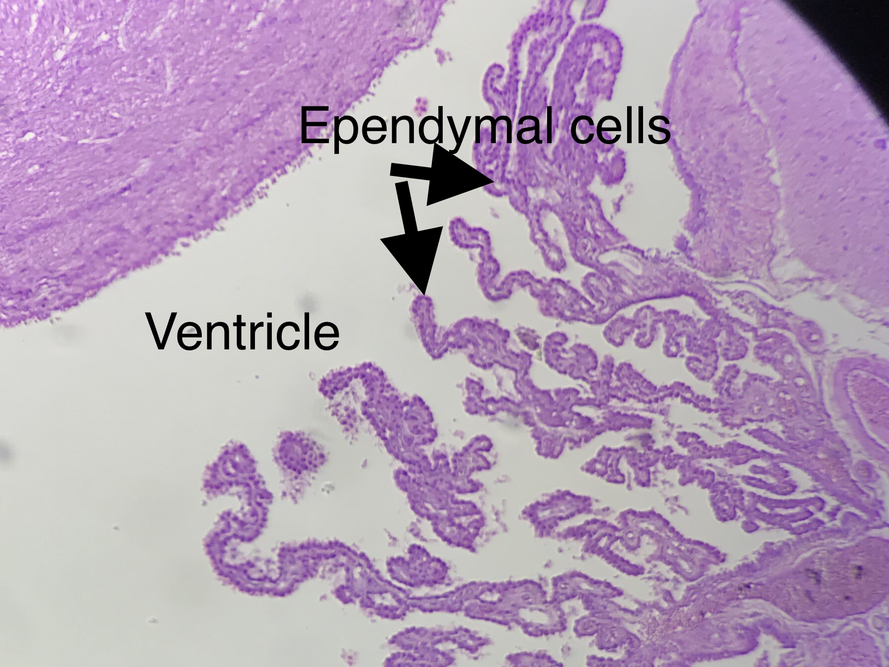
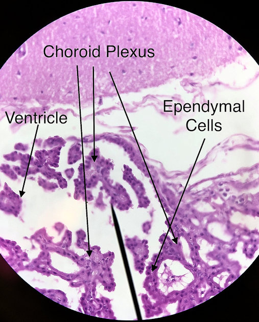
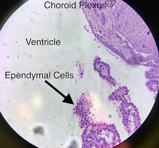
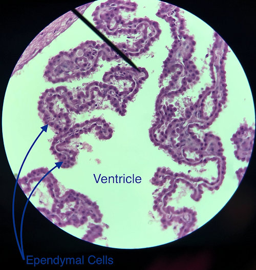
The histology on this page is of the 4th ventricle. It shows the choroid plexus and the ependymal cell of the plexus. The picture to the left is from the lab book. The picture on the right are student files from the fall of 2019.
Ependymal cell are found in brain structures called ventricles and produce a substance called cerebral spinal fluid (CSF). CSF buffers the chemical environment of the brain as well as washes away waste products found in the brain. Ependymal cells resemble ciliated epithelial cells and are found wrapped around a series of capillaries in the ventricles called the choroid plexus.
Scroll through the student pictures using the arrows. Try to identify the neurons and the glial cells. Your teacher may have you either draw or screen shot and label.
| Lab Book Image | Student Images |
|---|---|
|
1 /6
High Power
2 / 6
High Power
3 / 6
High Power
4 / 6
High Power
5 / 6
High Power
6 / 6
High Power
|