1 /8
Scanning
2 / 8
Low Power
3 / 8
High Power
4 / 8
High Power
5 / 8
Low Power
6 /7
Scanning
7 / 8
Osmic Acid, Low Power
8 / 8
Osmic Acid High Power
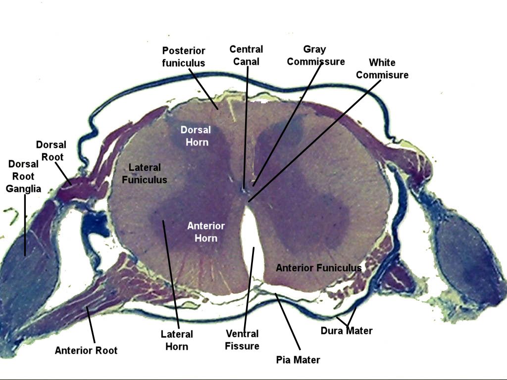
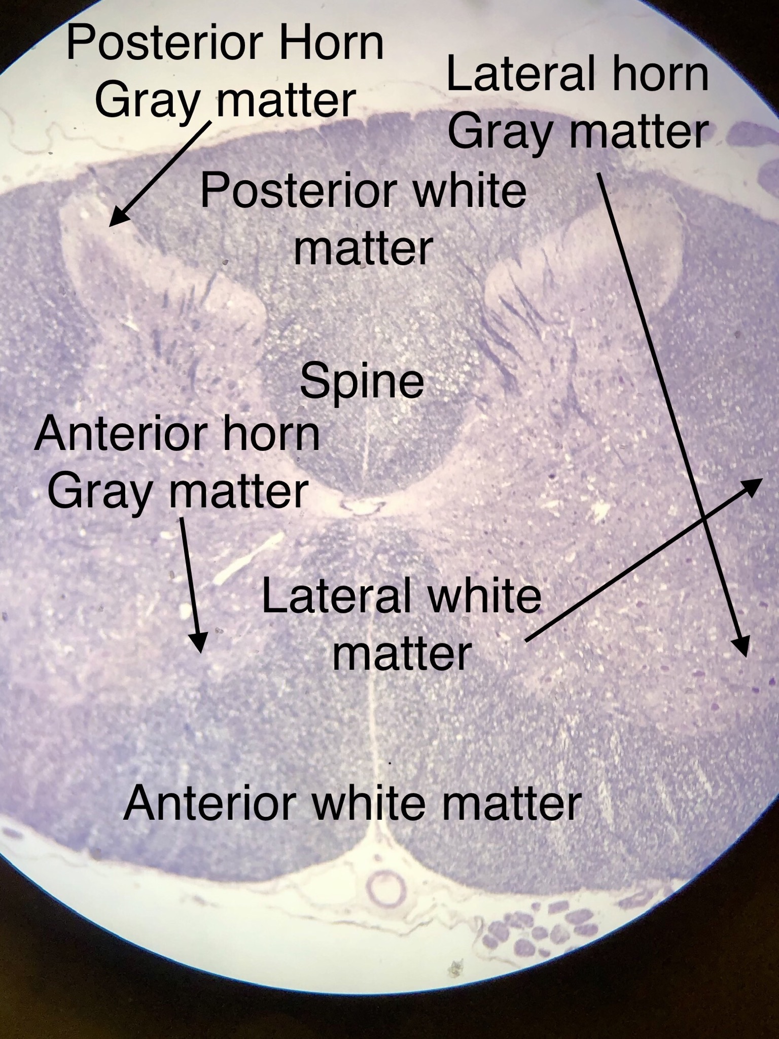
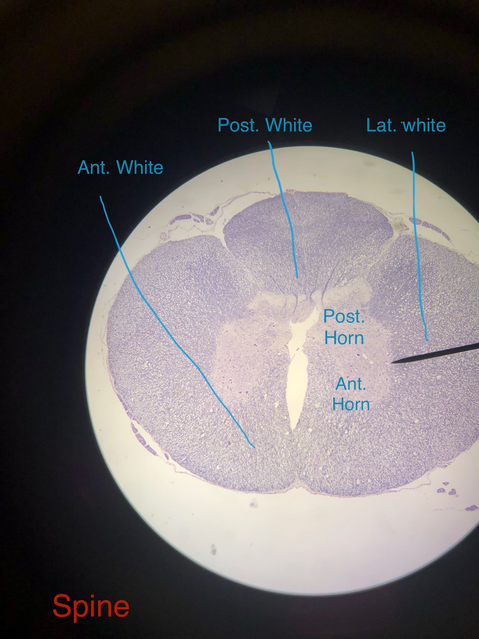
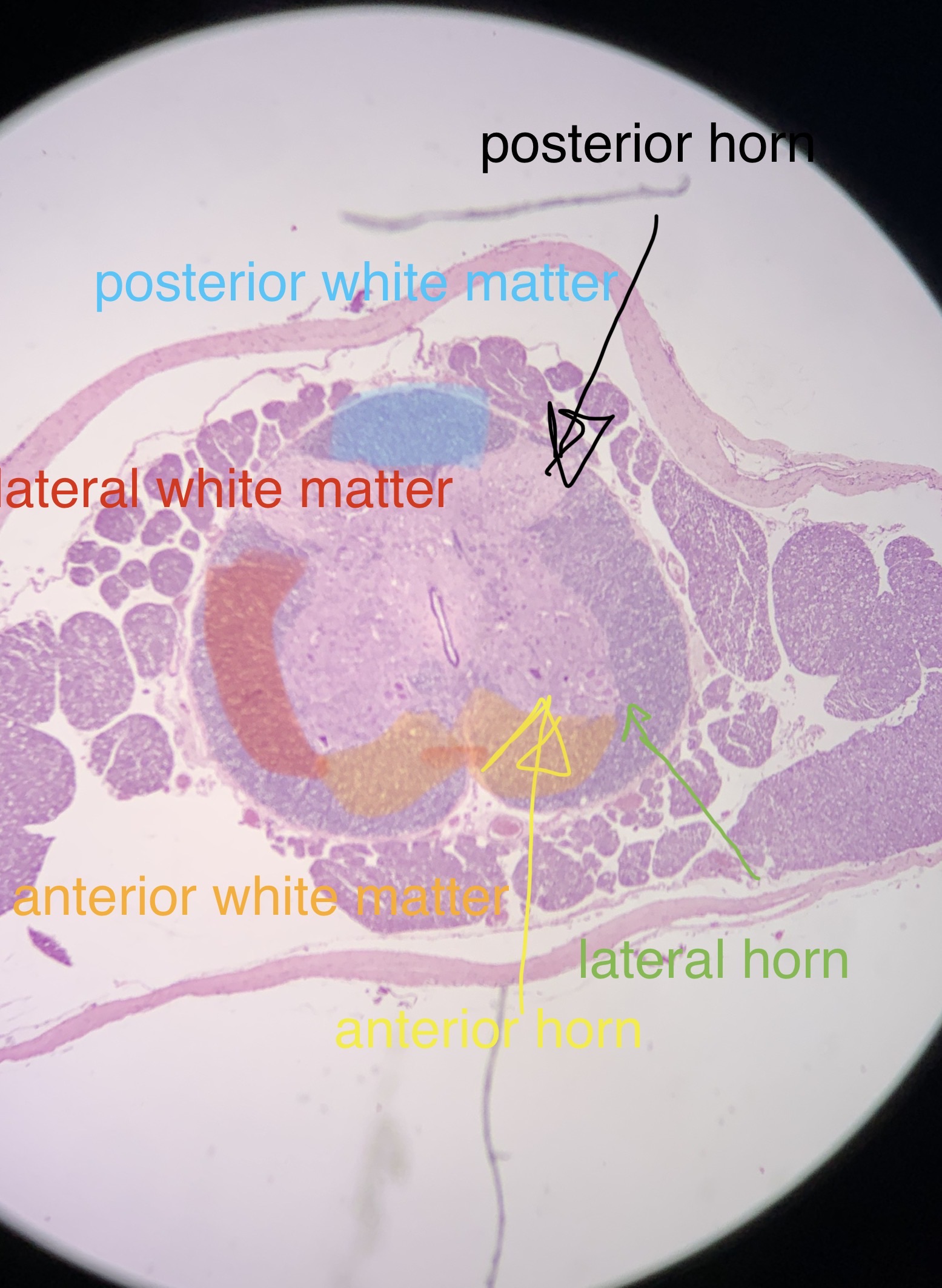
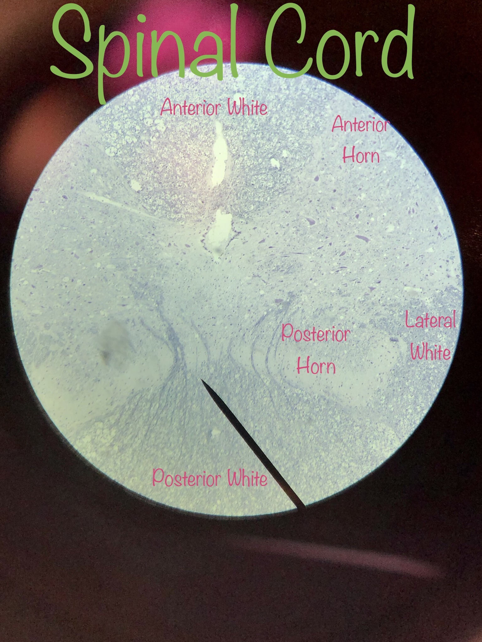
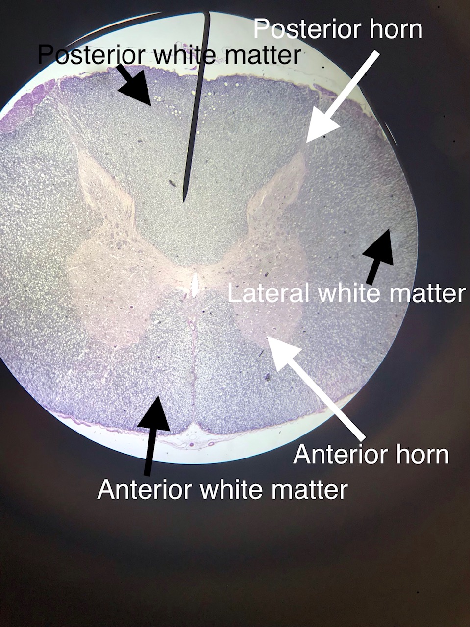
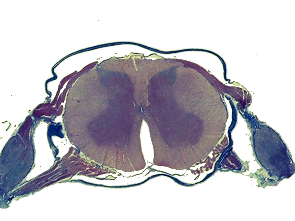
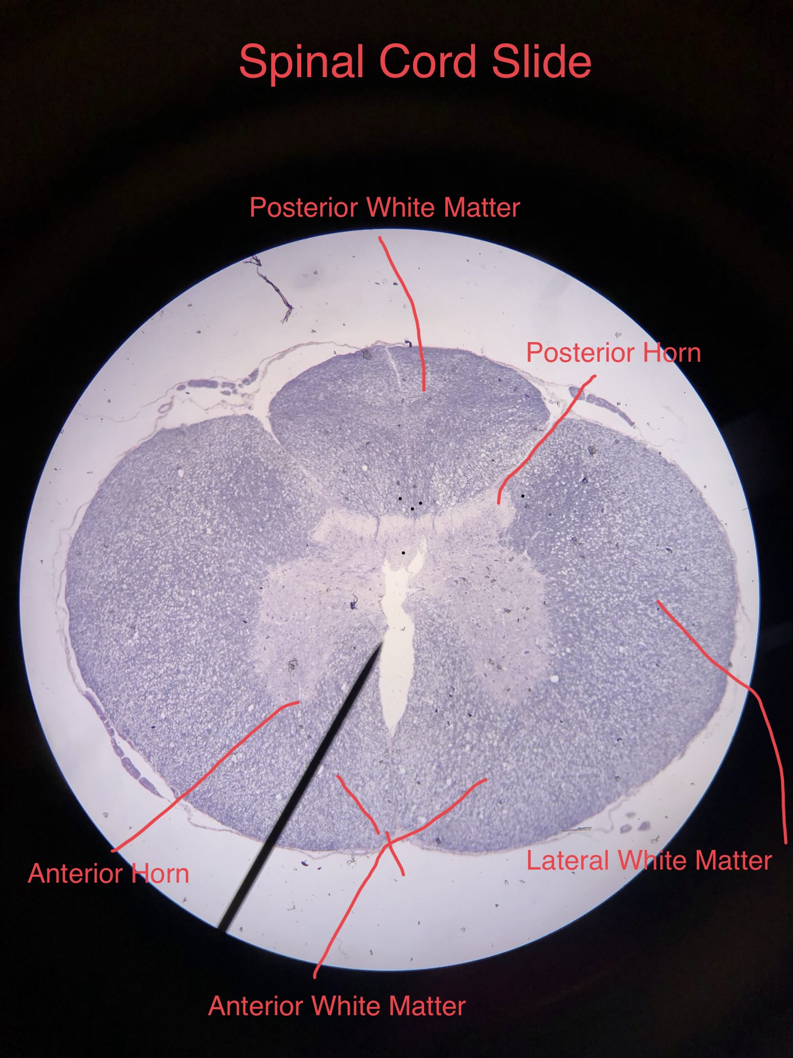
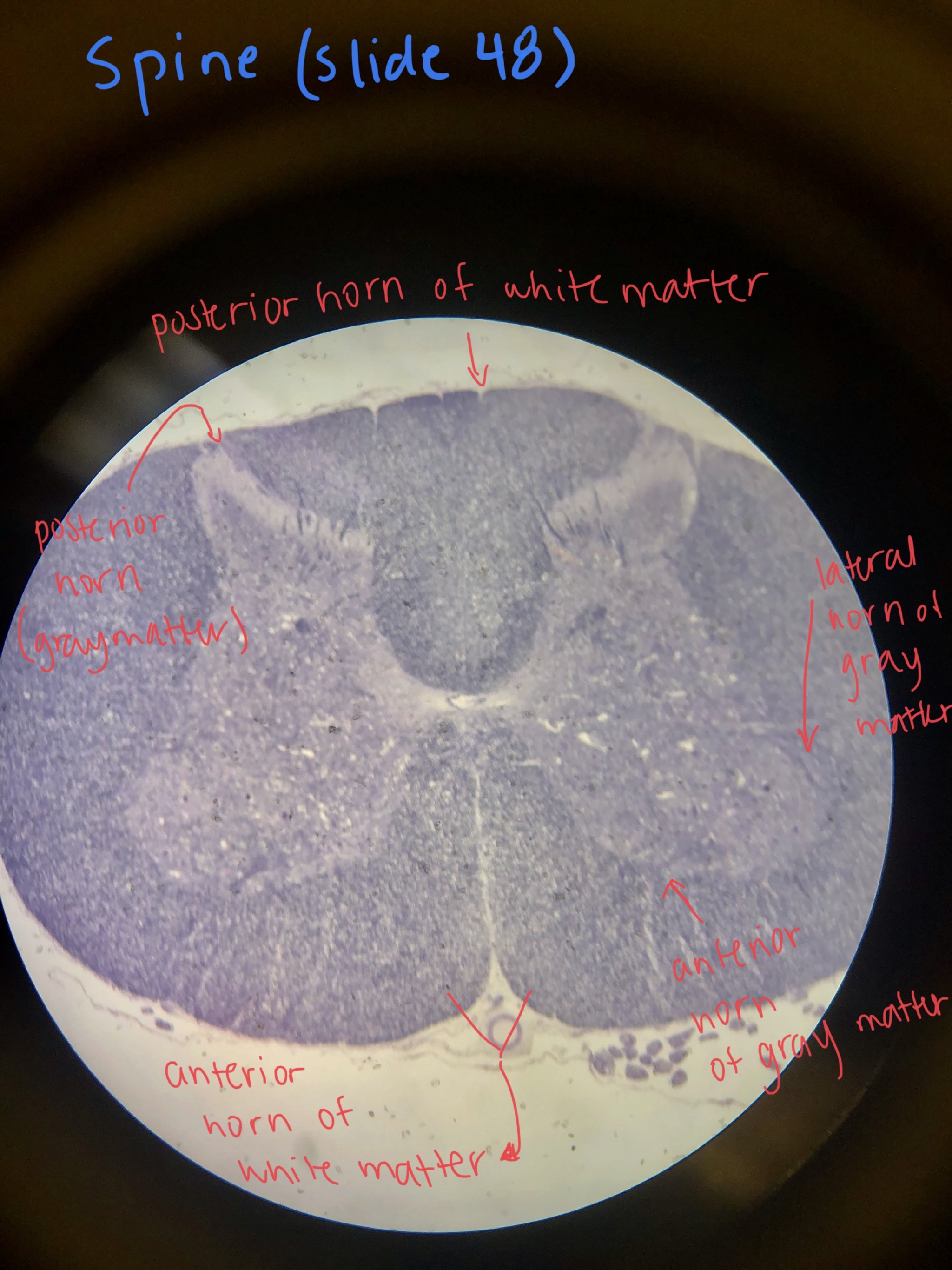
This page has histology of spine cross sections. It can be used to view parts of the spinal cord and do contrast gray and white matter. The picture to the left is from the lab book. The picture on the right are student files from the fall of 2019.
In the spine, things are reversed. The gray matter is central to the white matter and forms a butterfly shape with 3 identifiable horns. The posterior (dorsal) horns of the spine contain cell bodies of integration neurons. These will interoperate incoming signals. If a response is needed, these neurons send signals to the anterior horn as well as to the brain via the dorsal (posterior) white matter. The lateral and anterior (ventral) horns contain cell bodies of motor neurons that will leave the spine. These receive signals from the brain via the anterior and lateral white matter tracts. Since the gray matter is surrounded by white matter, the cell bodies in the spine are all considered parts of nuclei.
Scroll through the student pictures using the arrows. Try to identify the structures on the spine cross sections. Your teacher may have you either draw or screen shot and label.
| Lab Book Image | Student Images |
|---|---|
|
|
1 /8
Scanning
2 / 8
Low Power
3 / 8
High Power
4 / 8
High Power
5 / 8
Low Power
6 /7
Scanning
7 / 8
Osmic Acid, Low Power
8 / 8
Osmic Acid High Power
|