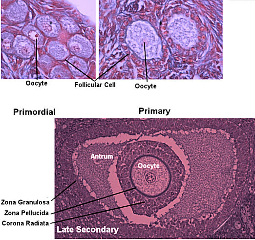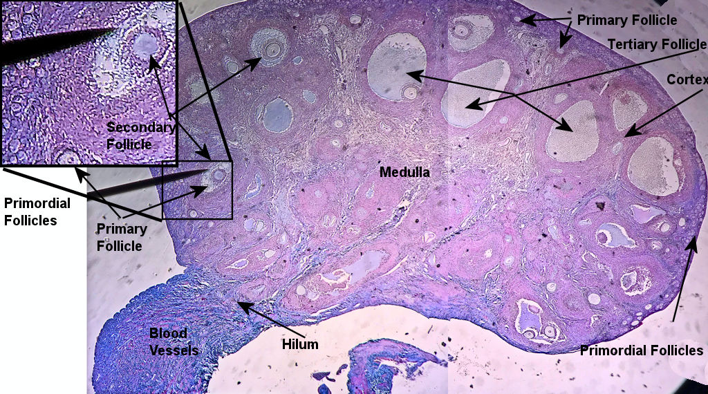

Histology of the ovary The ovary is divided into the cortex, which contains structures called follicles, and the medulla which contains blood vessels and nerves. Towards the outside of the cortex are primitive follicles called primordial follicles. The primordial follicles are made up of a layer of simple squamous supporting cells surrounding a primary oocyte that is suspended in the metaphase of its first meiotic division. Each month, follicle stimulating hormone causes several follicles to begin to develop. These follicles become primary follicles. The supporting cells become cuboidal and start to secrete messengers to aid in the development of the oocyte, the follicle, and the functional endometrium. A capsule called the zona pellucida forms around the oocyte as well. The zona pellucida will be ovulated with the oocyte and contains receptors and enzymes that aid the sperm in penetrating the oocyte. The supporting cells begin to increase in number forming 2 layers of cuboidal cells. A fluid fill cavity called an antrum starts to form between the supporting cells and the follicle. This marks the follicles progression from a primary to a secondary follicle. A second layer of cuboidal cells called the corona radiate form around the zona pellucida and the fluid in the antrum now fully separates the oocyte from the supporting cells. The follicle is now considered a tertiary or vesicular follicle and the oocyte is about to be released. After ovulation, the supporting cells become a structure known as the corpus luteum. The corpus luteum makes progesterone that maintains the functional endometrium for a week or so after ovulation and the corpus luteum is identifiable by its yellowish color. When the corpus luteum stops making progesterone, it begins to atrophy and becomes white in color. This structure is known as the corpus albicans. The corpus albicans will eventually become connective tissue and that gets reabsorbed.
These pictures were taken by me in the spring of 2021. The first 2 unlabeled pictures show the whole ovary. Then pictures 3-8 show different follicle stages. The pointer is pointing to the yellow label on the botton. This model what the ovary looks like.
| Follicles Image | Unlabeled Images |
|---|---|

|
|
| Whole Ovary Labeled | |

| |