Respiratory Histology | Histology Home Page | Site Home Page
Lung
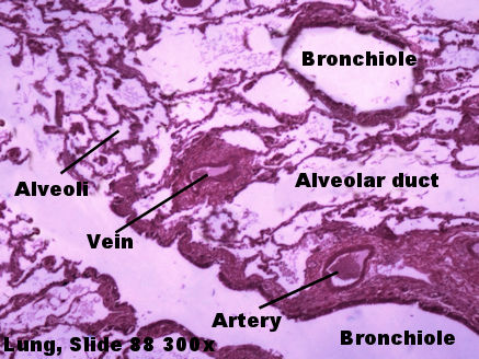
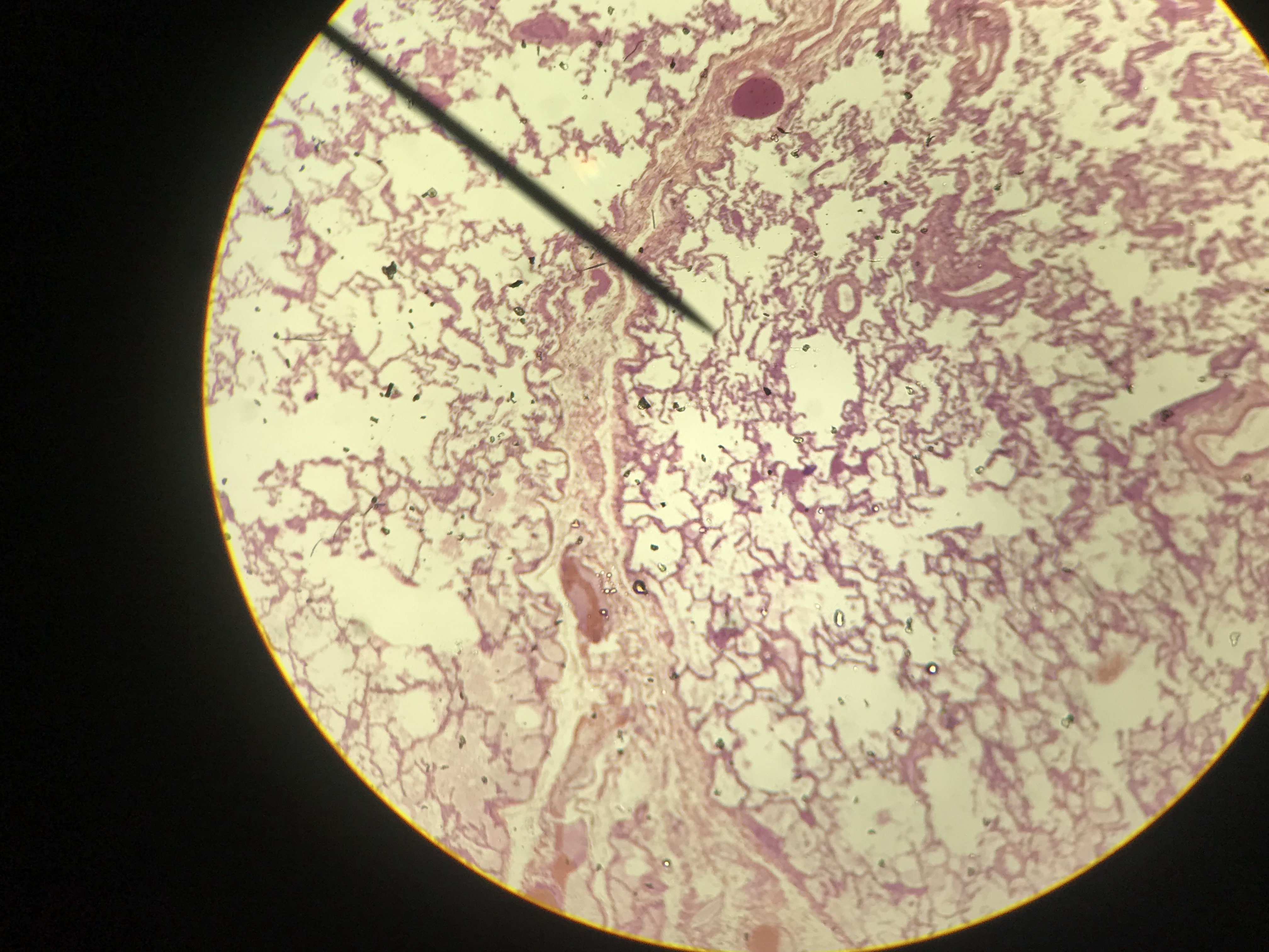
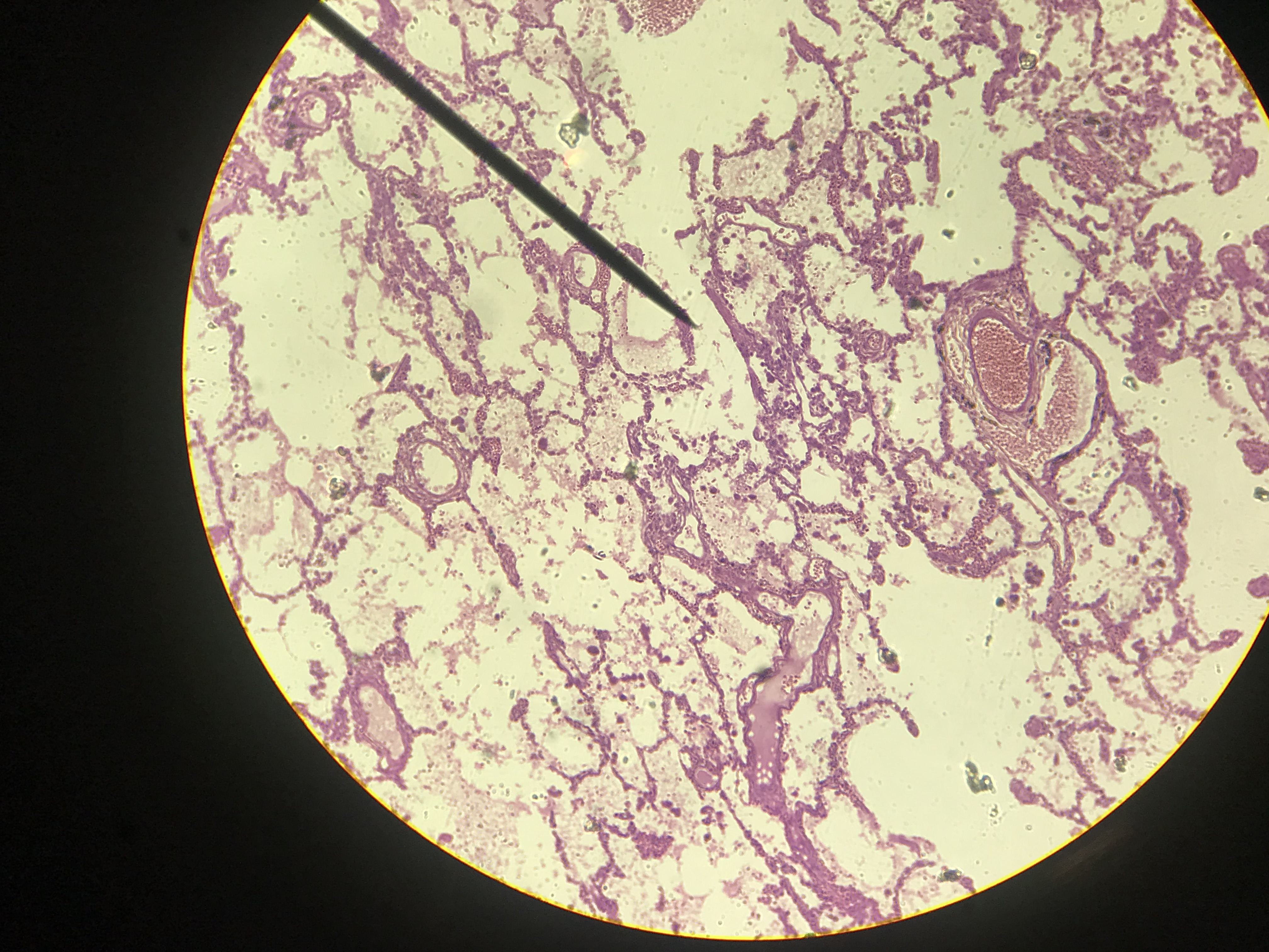
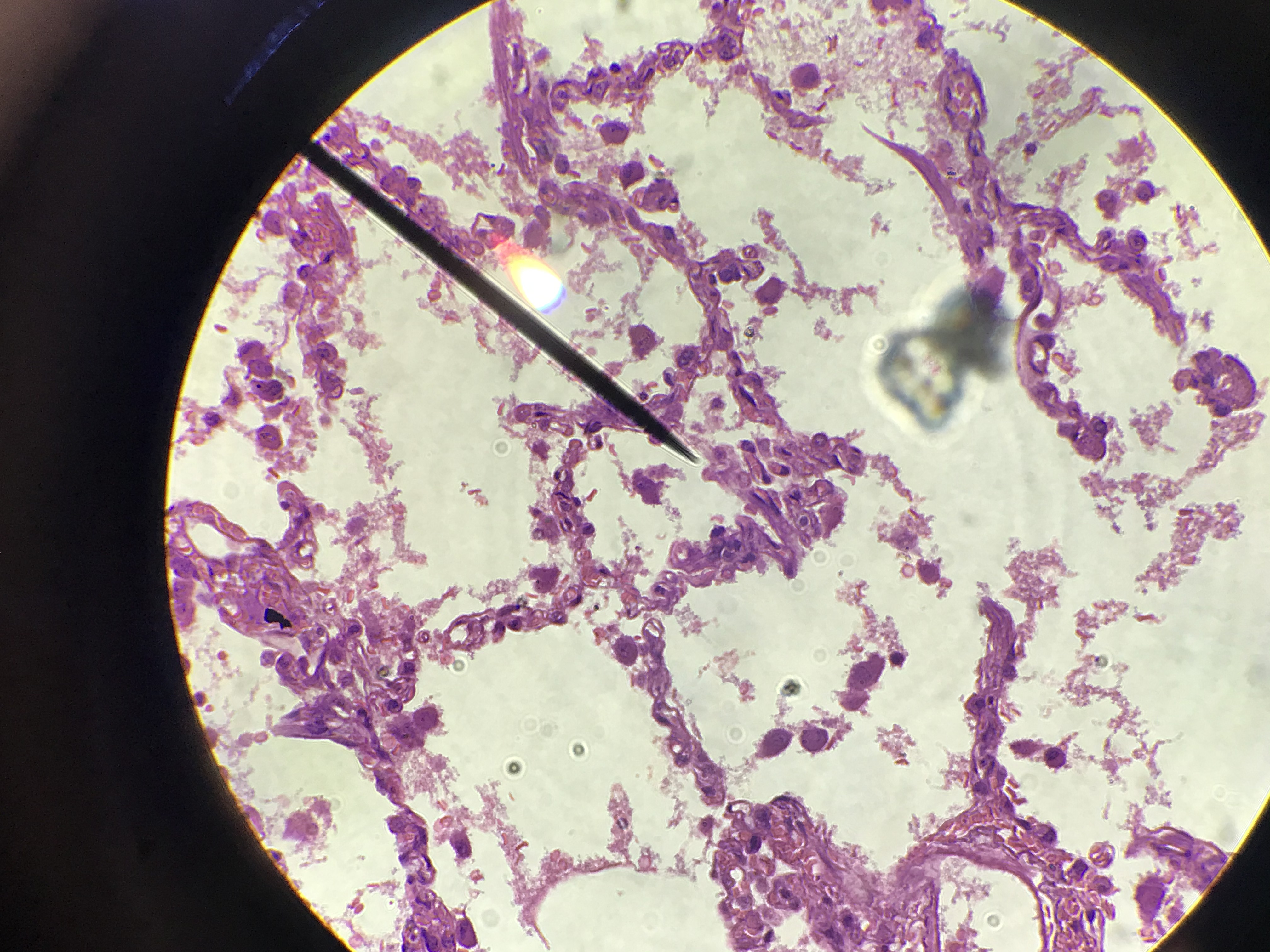
Observe the lung slide. Try to tell the difference between the following
- Alveolar sacks lined with simple squamous epithelial and lacks a submucosa
- Alveolar ducts lined with simple squamous epithelial
- Bronchioles lined with simple cuboidal epithelial and a submucosal of smooth muscle.
- Arteries, arterioles, venules, and veins. On capillaries too.
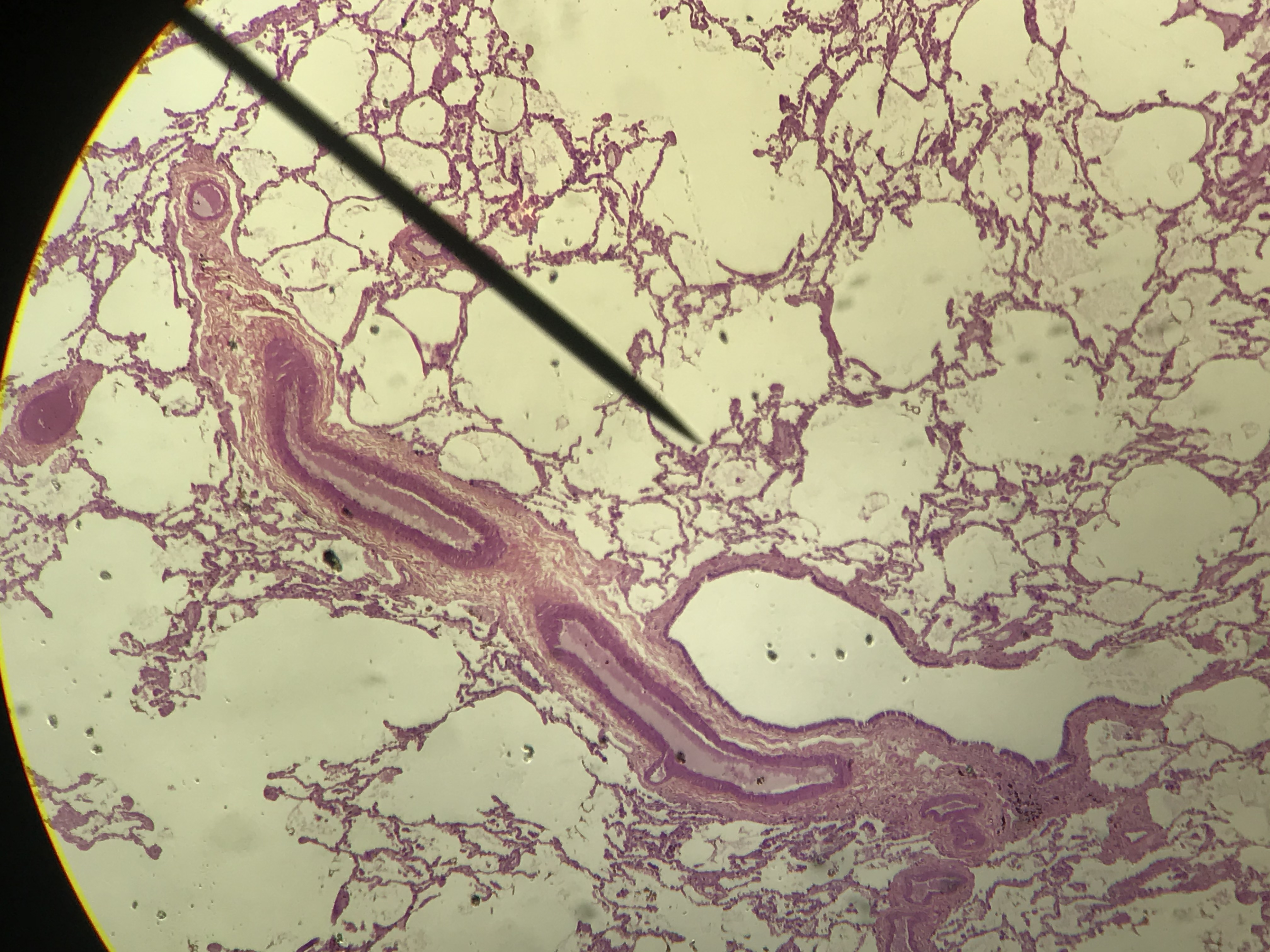
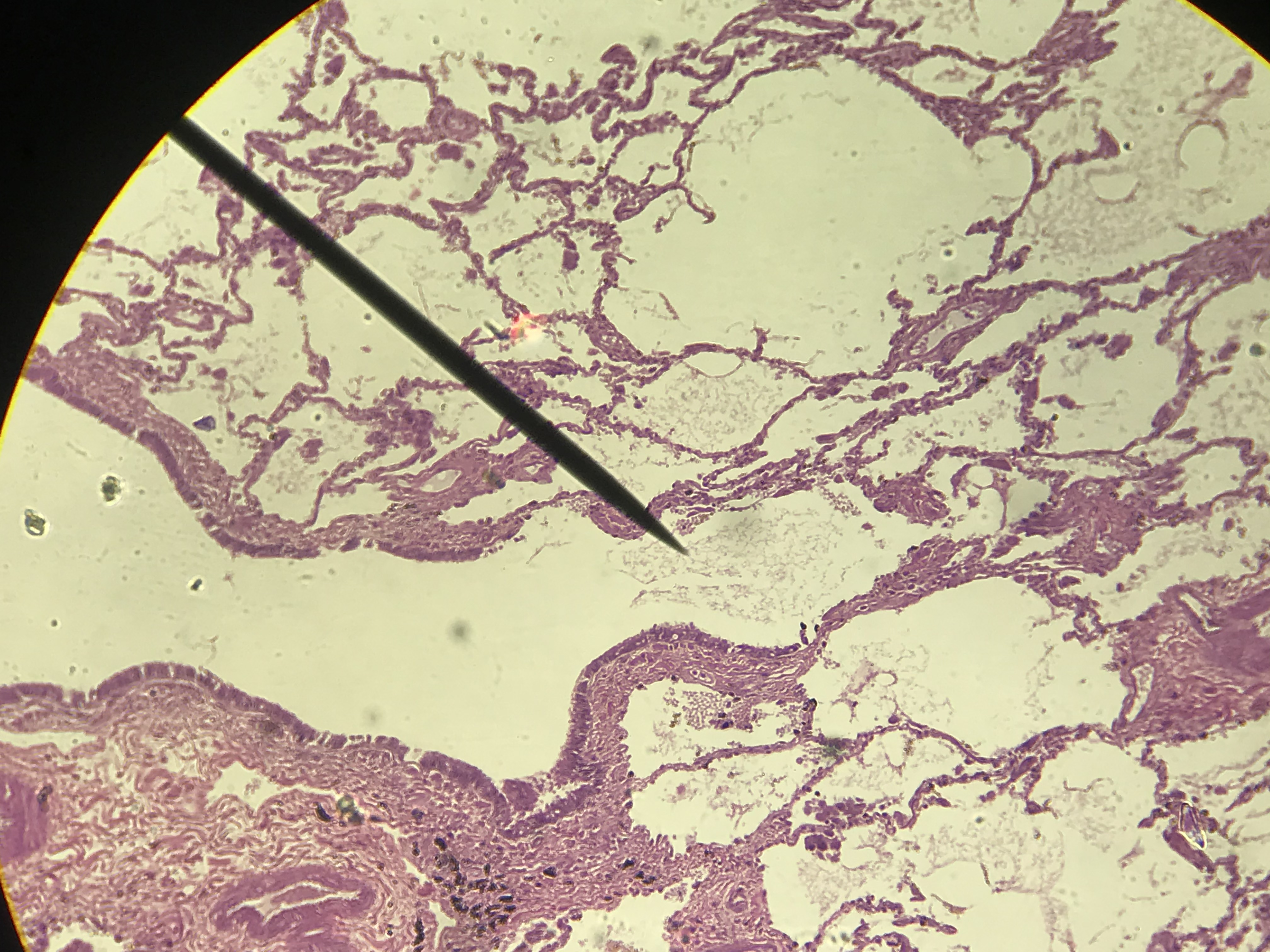
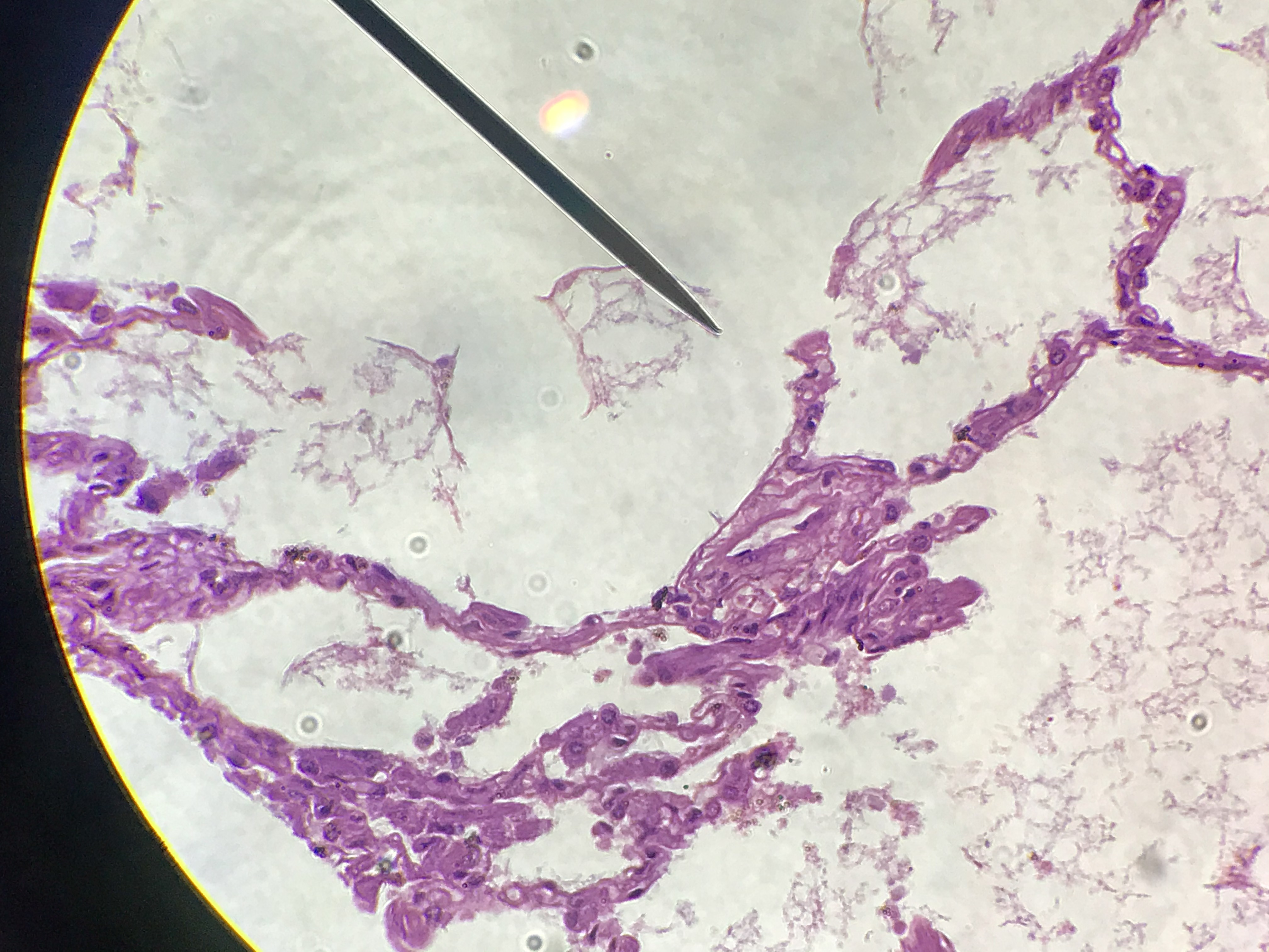
These pictures were taken by me in the spring of 2021. They progress from scanning power (40x) to high power (400x).
Go through the pictures. Select one, draw it, and label the layers. Note, you can right click/command click on the pictures and open them in new windows. This will enlarge them.
| Labeled Image |
Unlabeled Images |
 |
s
1/6
scanning
2/6
low
3/6
high Power
4/6
scanning Power
5/6
low Power
6/6
high Power
❮
❯
|