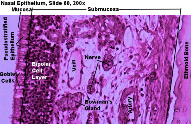
The nasal cavity is lined with ciliated pseudostratified epithelial tissue with mucus producing goblet cells. The mucus moistens the tract and traps foreign particles that the cilia sweep out. Because this layer secretes mucus in is called the mucosal layer. Under the mucosal layer is the submucosal layer which is composed of loose connective tissue (typically areolar), blood vessels, nerves and occasionally submucosal glands which often secrete something into the mucosal layer. In the nasal cavity, the nuclei of bipolar cells can be observed right under the epithelial tissue. In addition mucus producing glands can be visable. Look out for arteries and veins in this layer as well. Deep to the submocusal layer will be either the ethmoid bone or cartilage.
These pictures were taken by me in the spring of 2021. They progress from scanning power (40x) to high power (400x). Go through the pictures. Select one, draw it, and label the layers. Note, you can right click/command click on the pictures and open them in new windows. This will enlarge them.
| Labeled Image | Unlabeled Images |
|---|---|
 |
|