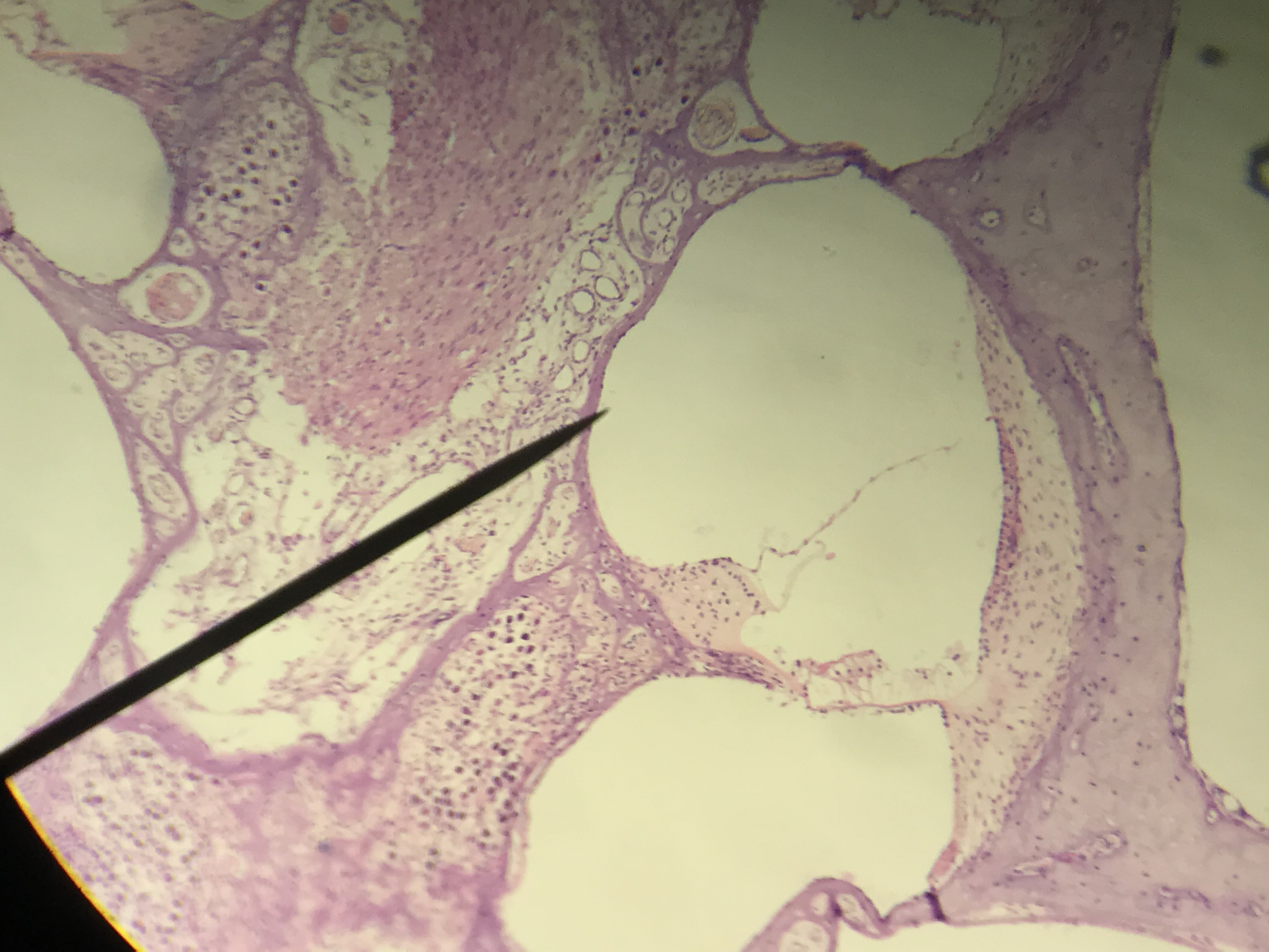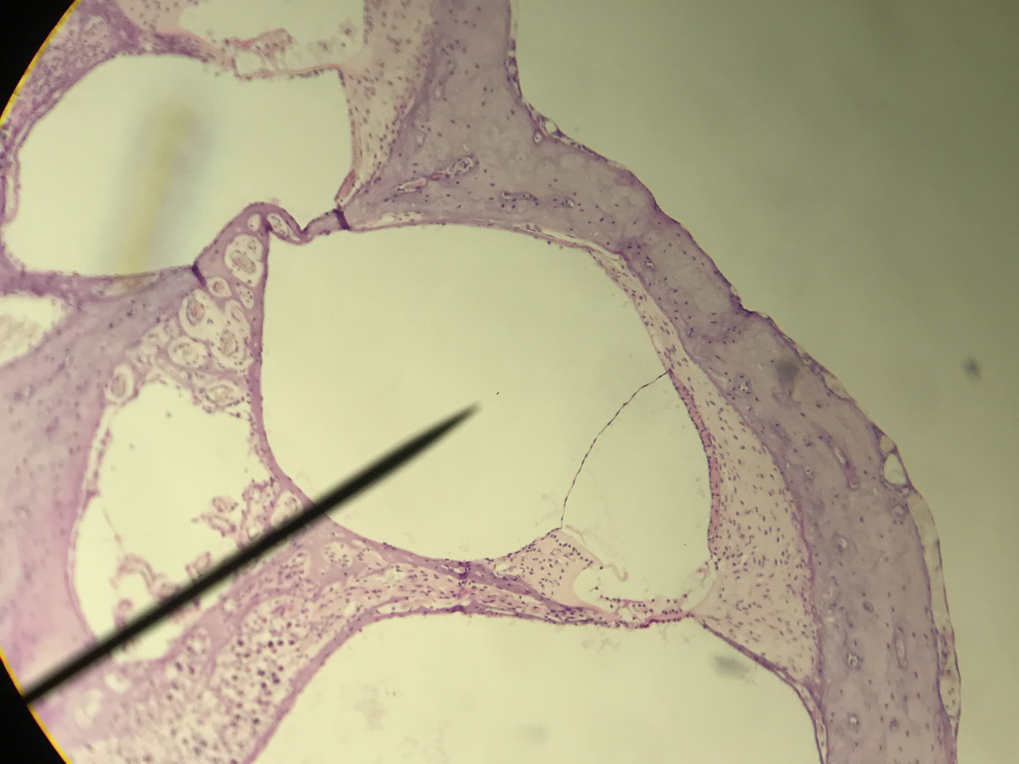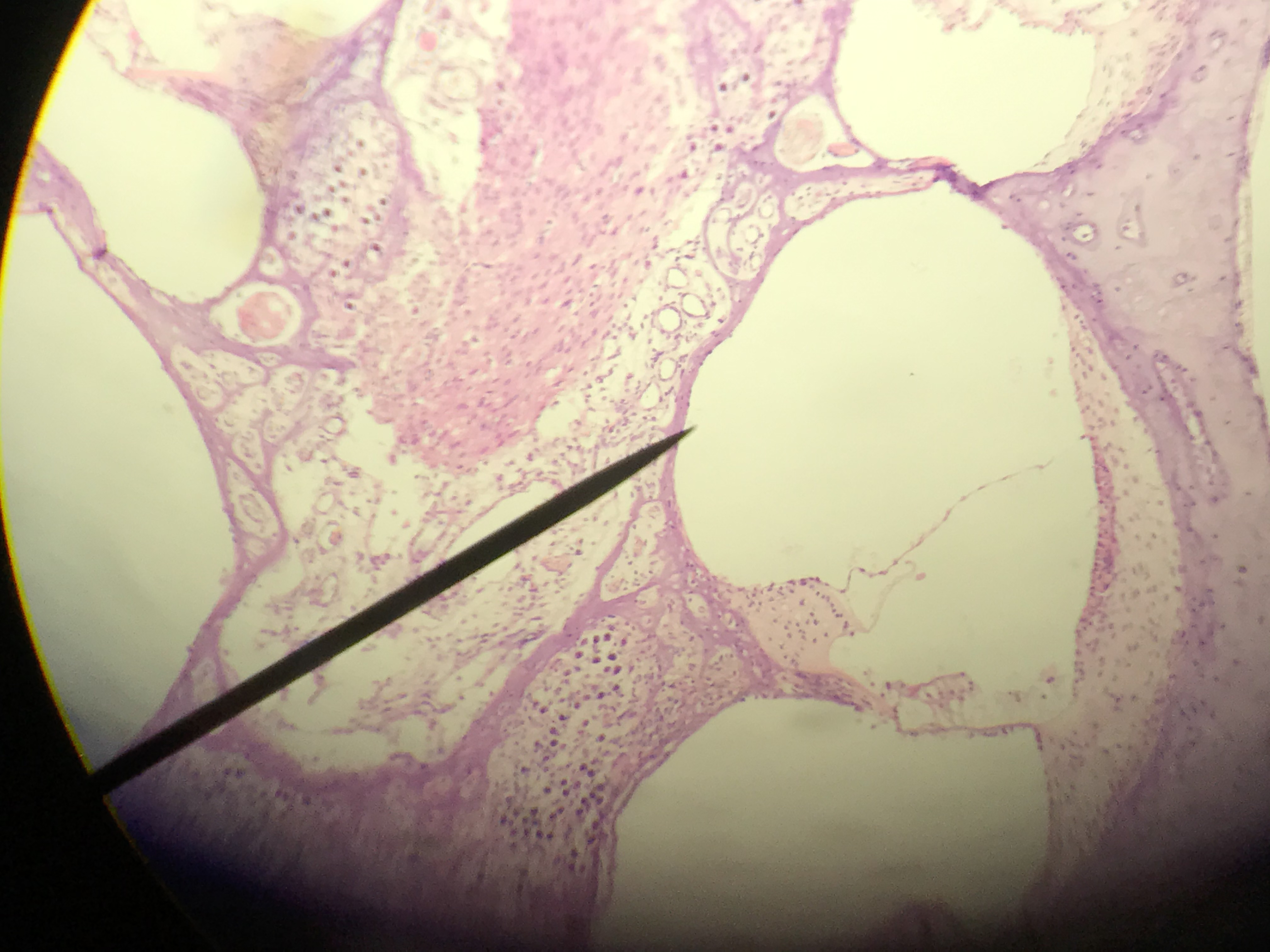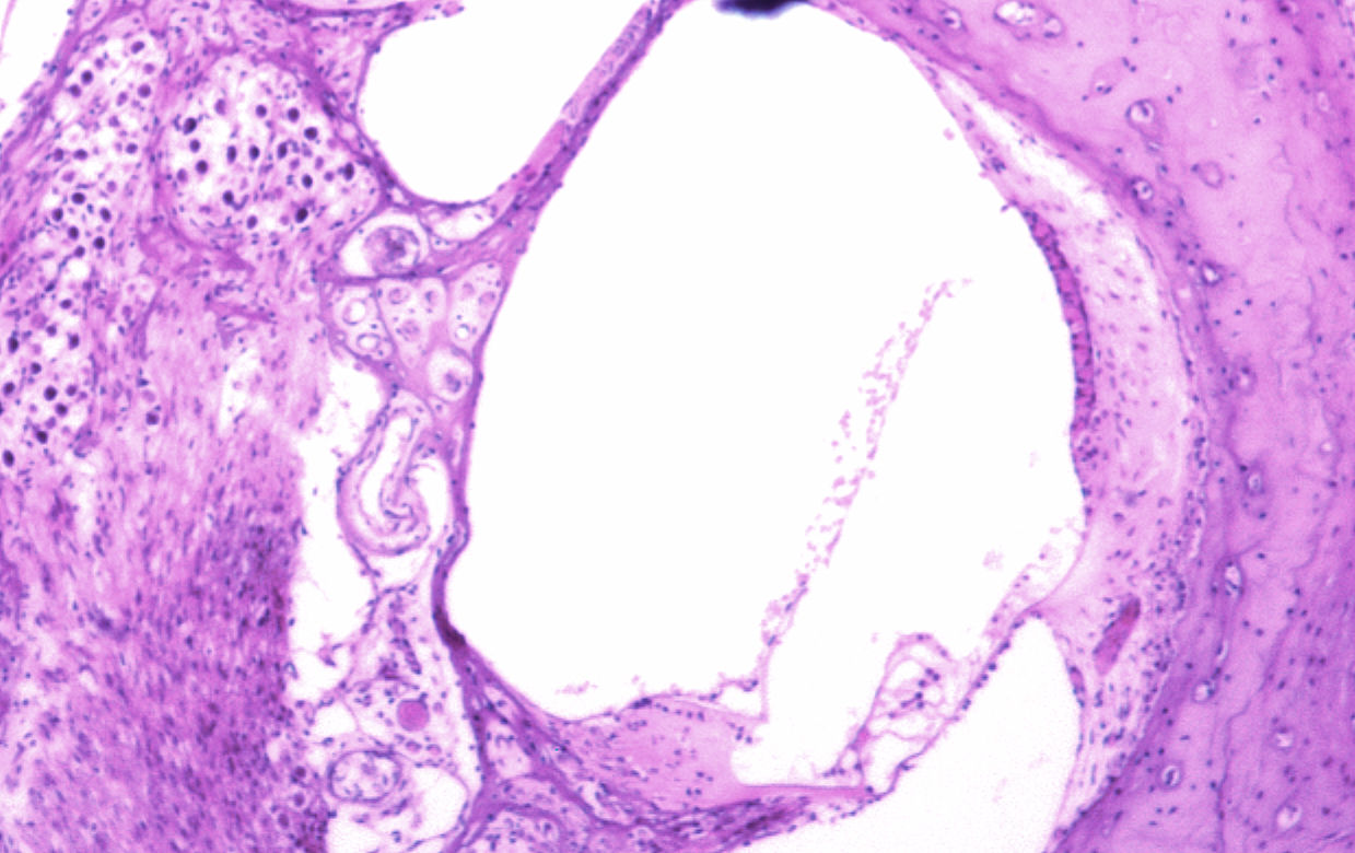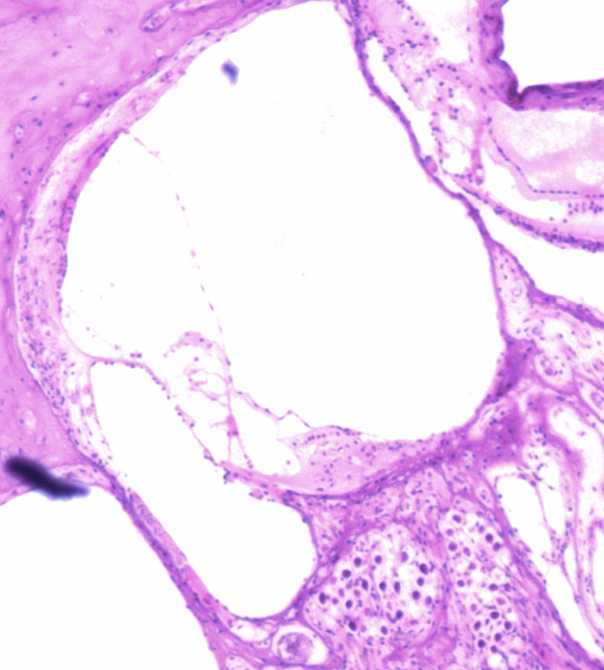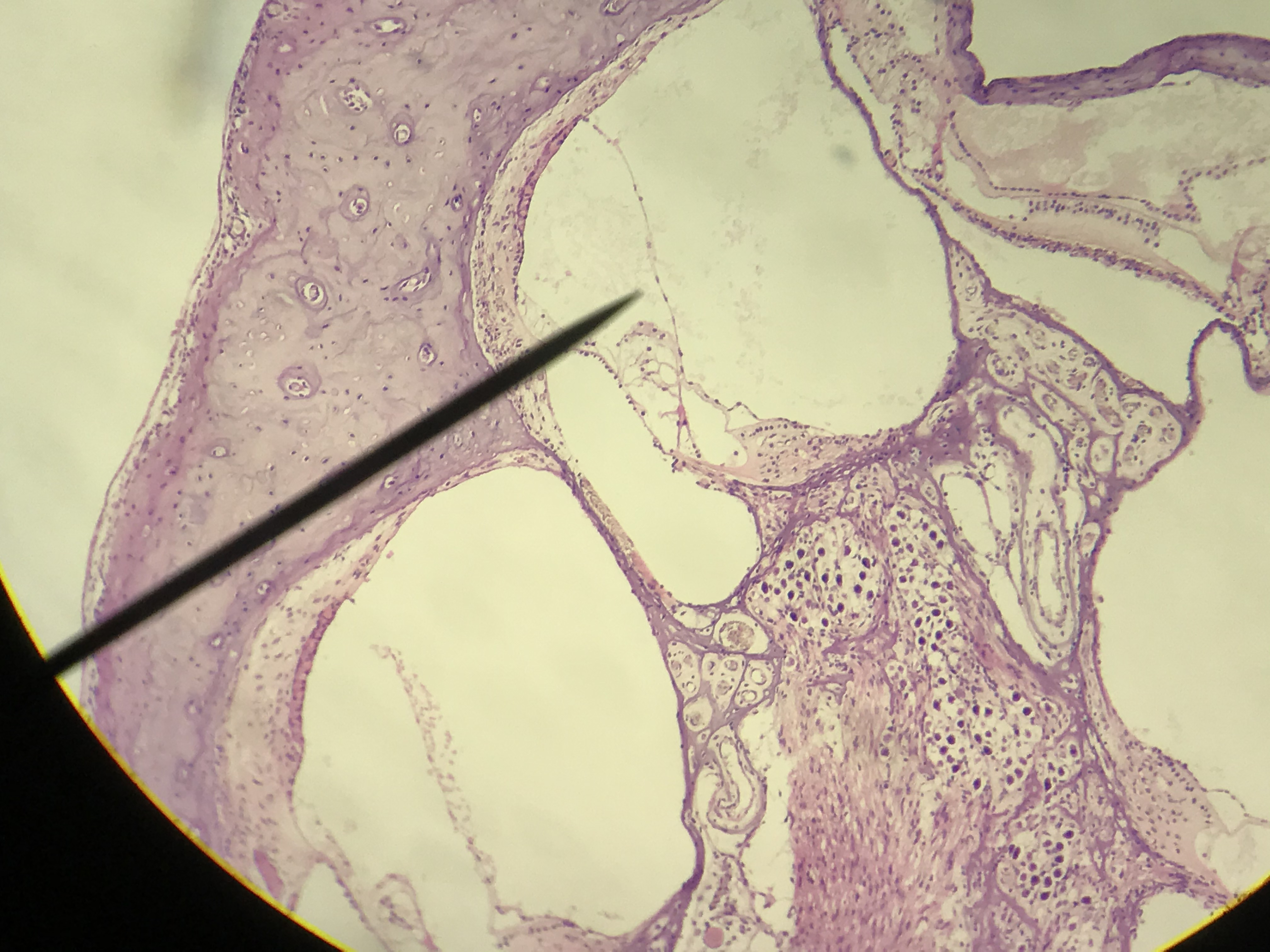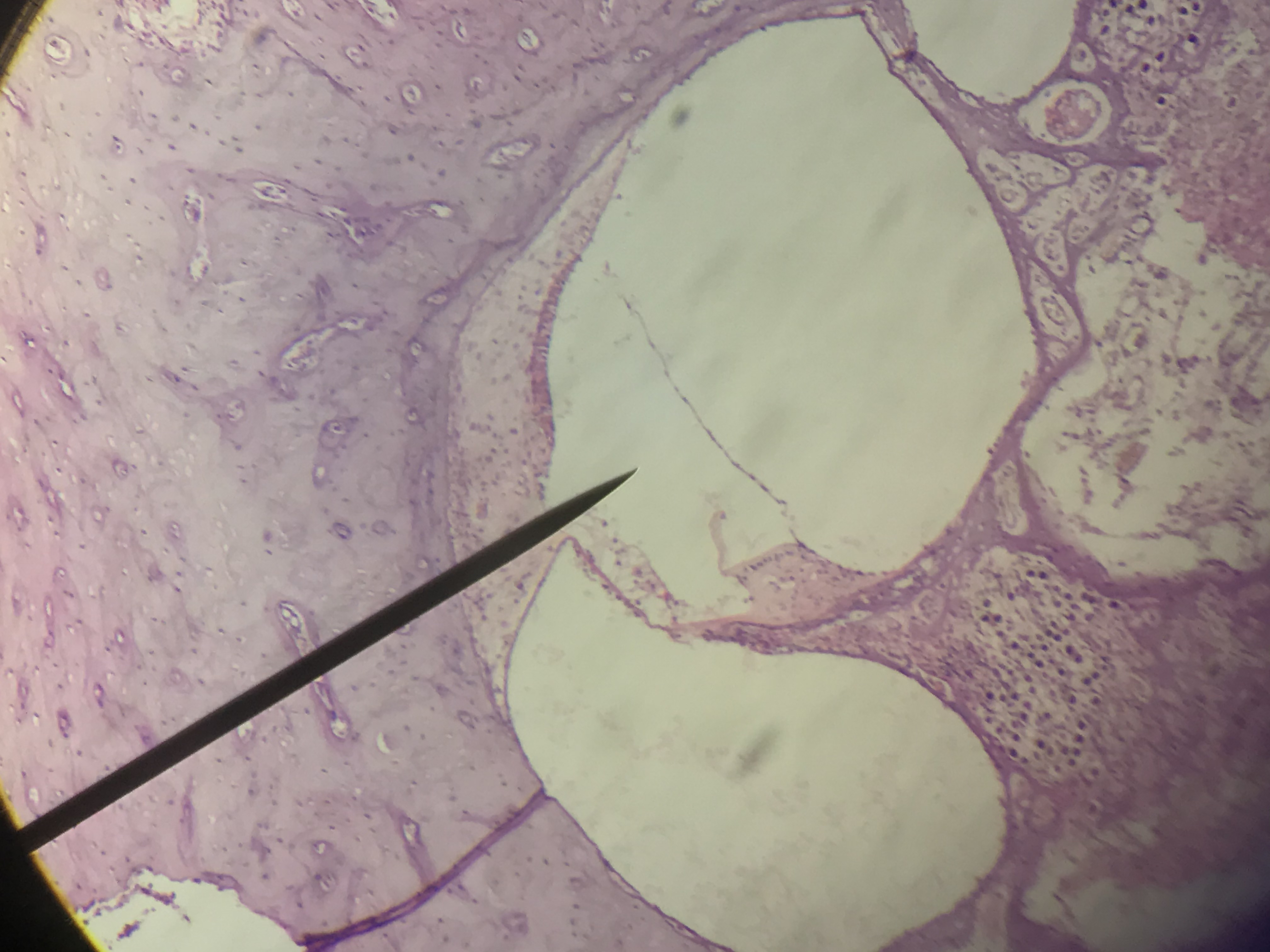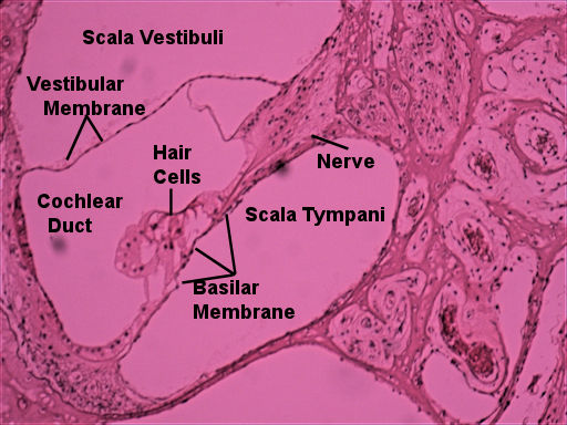
This page has histology of the cochlea.above the cochlea has three ducts that travel down its length. The cochlear duct is in the center and it is bordered by 2 membranes. The thin vestibular membrane is on one side while the basilar membrane, which the hair cells sit on, is on the other. On the other side of the vestibular membrane is the scala vestibuli (vestibular duct) which extends from the oval window. When the stapes pushes on the oval window, the vibrations disrupt the perilymph in the scala vestibuli. The waves from the disruption strikes the vestibular membrane which disrupts the endolymph in the cochlear duct. This causes the tectorial membrane to push on the hair cells. Lastly, the scala tympani (tympanic duct) is below the basilar membrane and is actually a continuation of the scala vestibuli. The vibrations continue down the scala tympani and eventually hit the round window.
The pictures were taken by me over various semesters. The lab book's picture is on the left and unlabeled picthers, which you can scroll through with the arrows, are on your right.Compare the unlabeled pictures with the labeld picture and try to find the structures from your lab book's picture. Then select one of the pictrues to draw in your lab book. This model is an enlarged view of what you are looking at.
| Lab Book Image | Student Images |
|---|---|
|
|
|