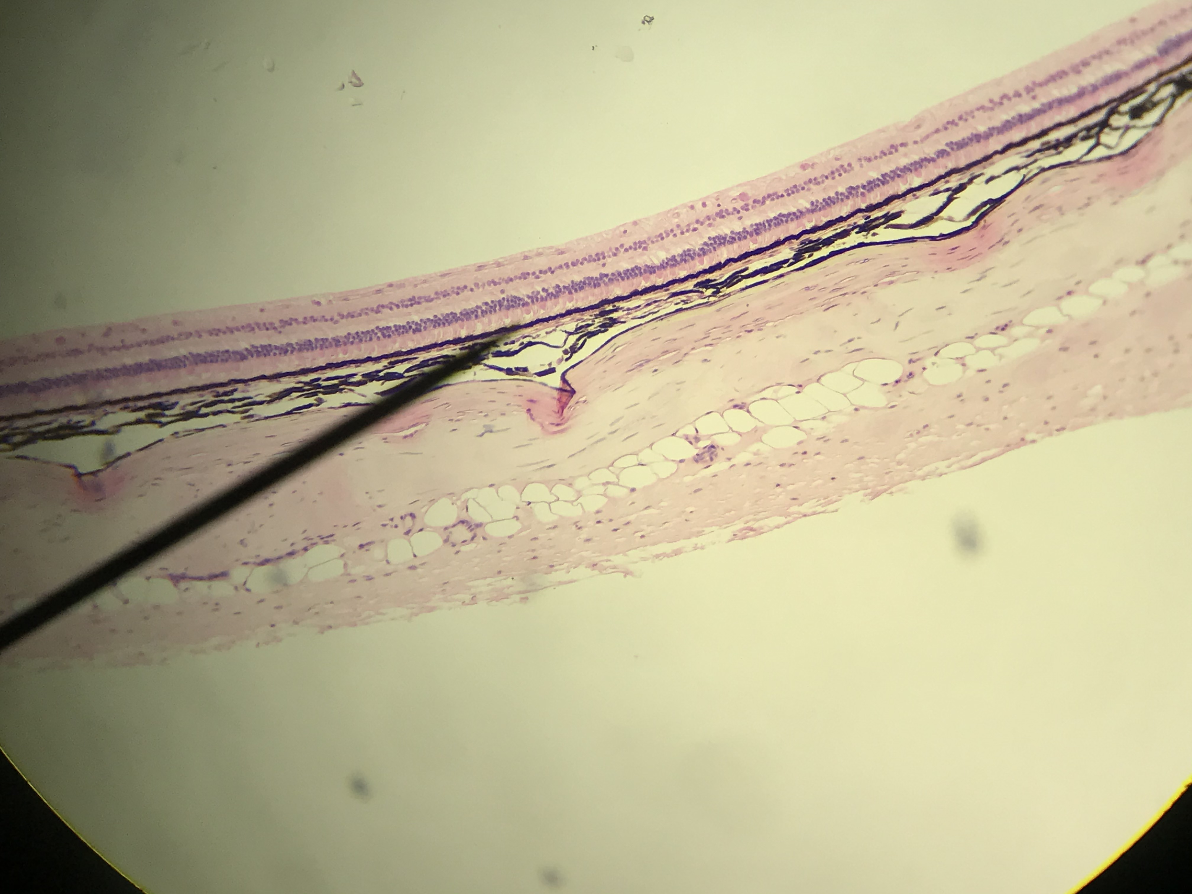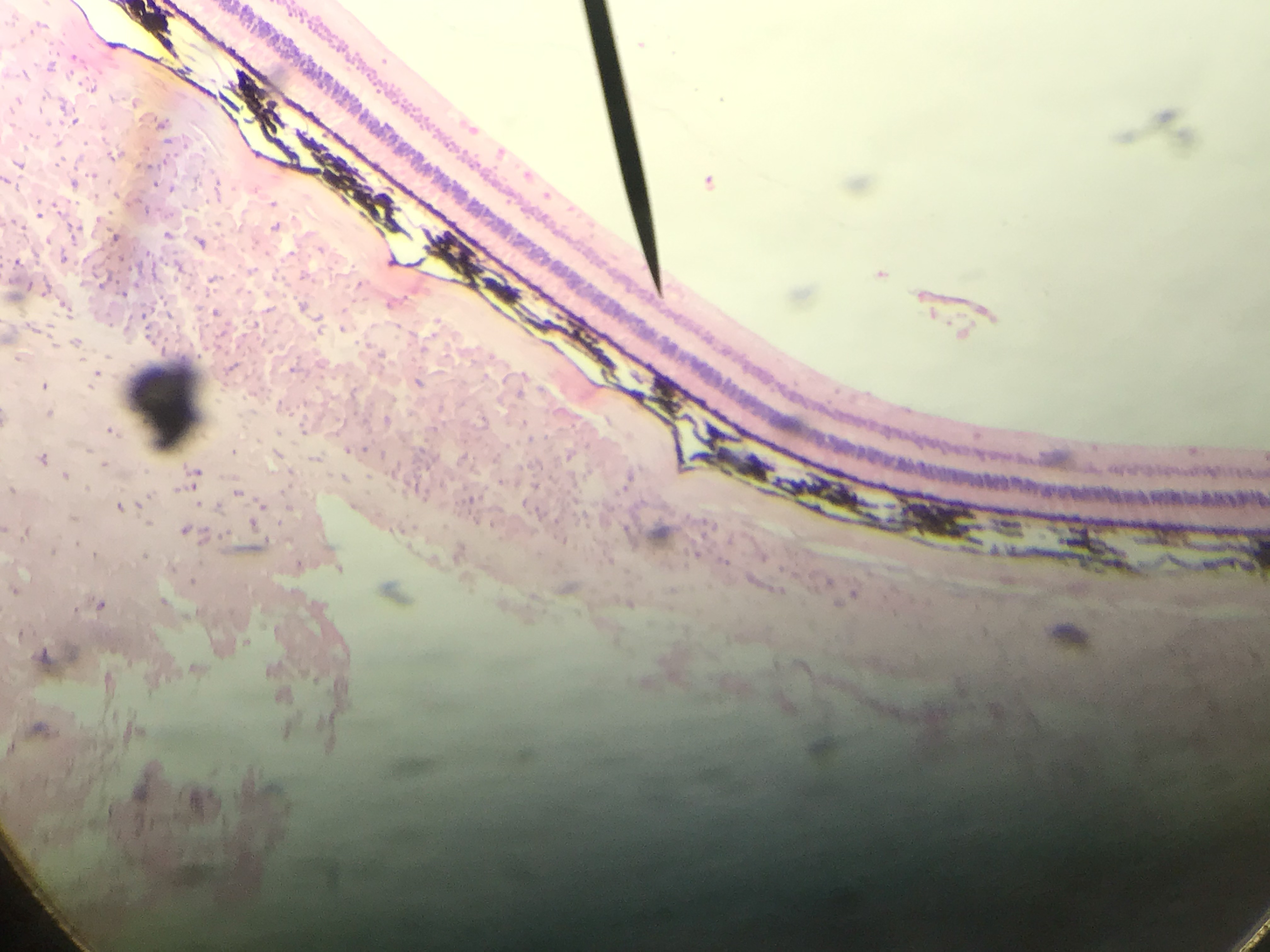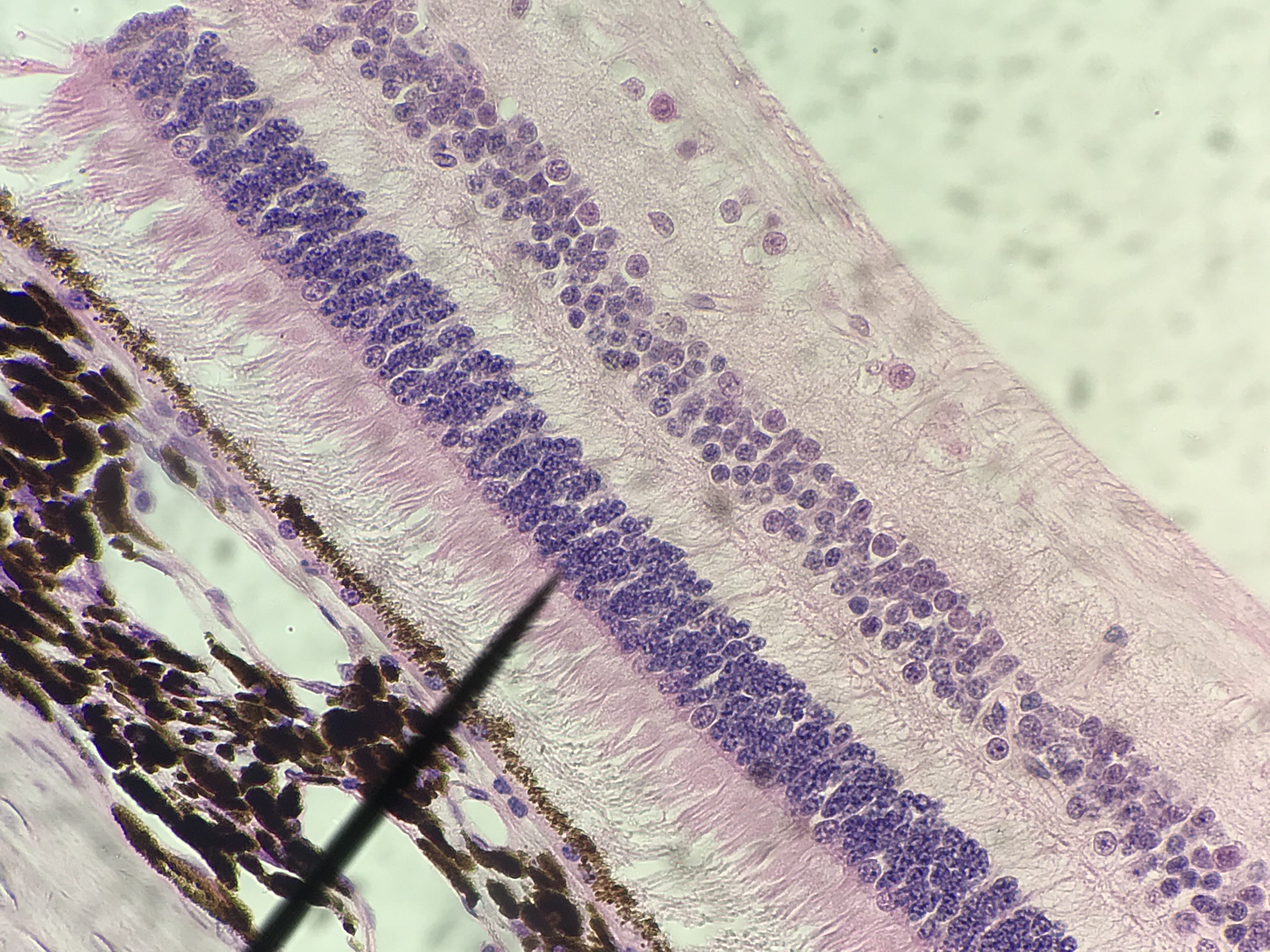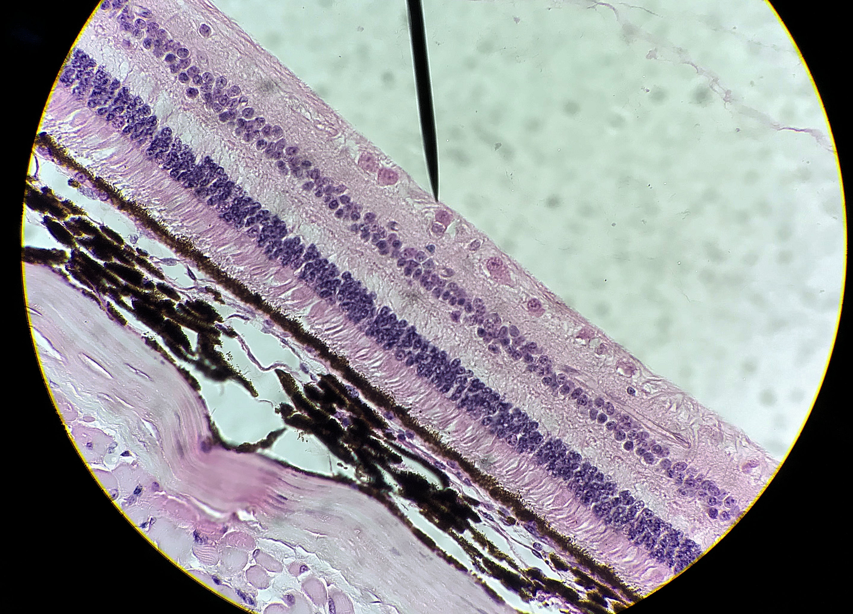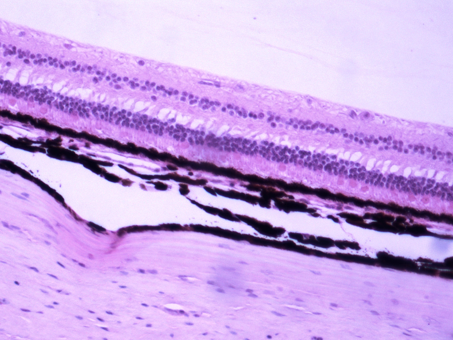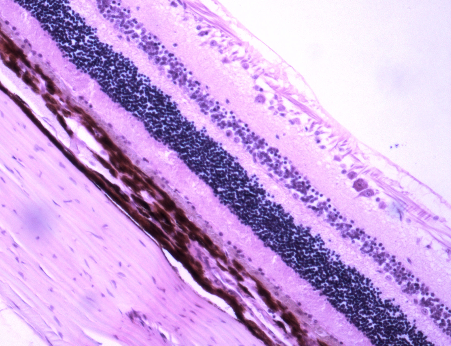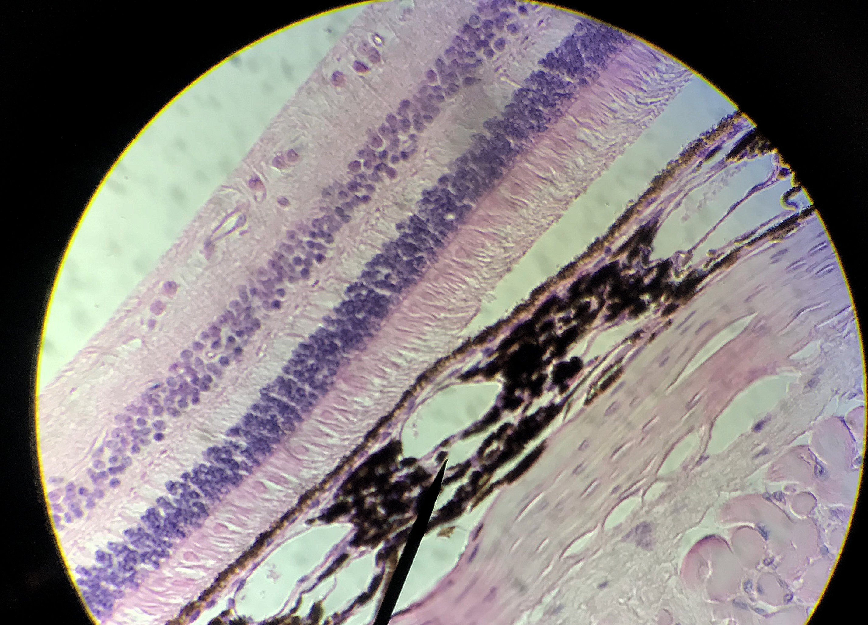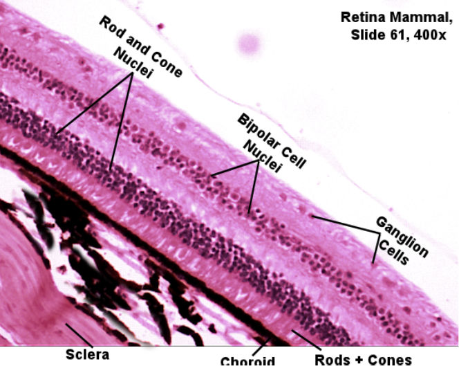
This page has histology of the retina. The retina has 3 layers of cells which help to convert what you "see" into nural impulses. Immediately adjacent to the black choroid you will see the rod and cone cells. Above the cells you will see the nuclei layer for the rods and cones. This should give you a good indication of how many photo receptors are in the retina. The bipolar cells may be hard to identify but you should be able to see the nuclei of the bipolar cells in a distinct layer. The last layer will be the nuclei of the ganglion cells.
The pictures were taken by me over various semesters. The lab book's picture is on the left and unlabeled picthers, which you can scroll through with the arrows, are on your right.Compare the unlabeled pictures with the labeld picture and try to find the structures from your lab book's picture. Then select one of the pictrues to draw in your lab book. This model is an enlarged view of what you are looking at.
| Lab Book Image | Student Images |
|---|---|
|
|
|