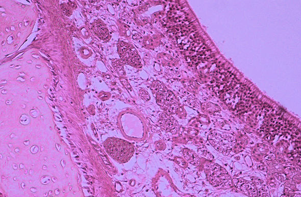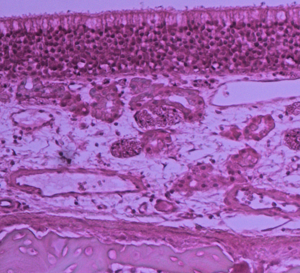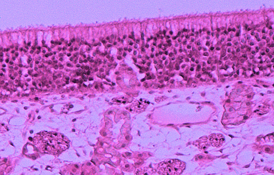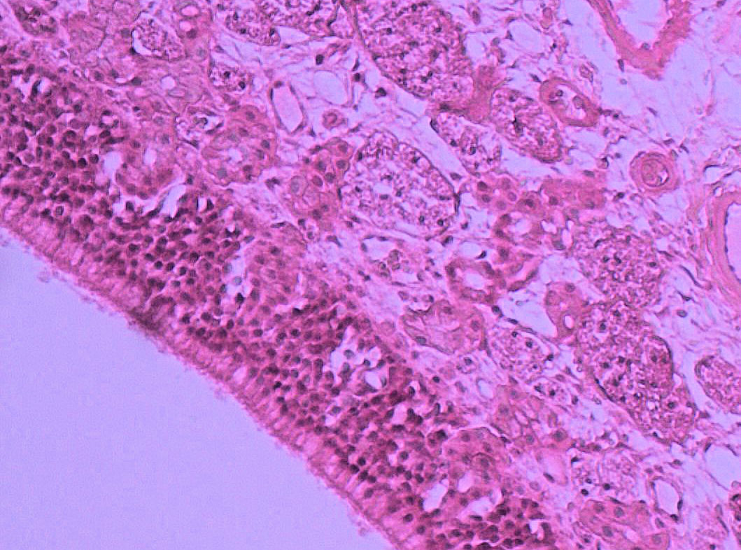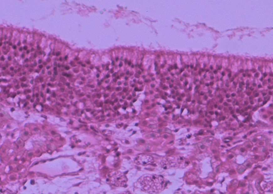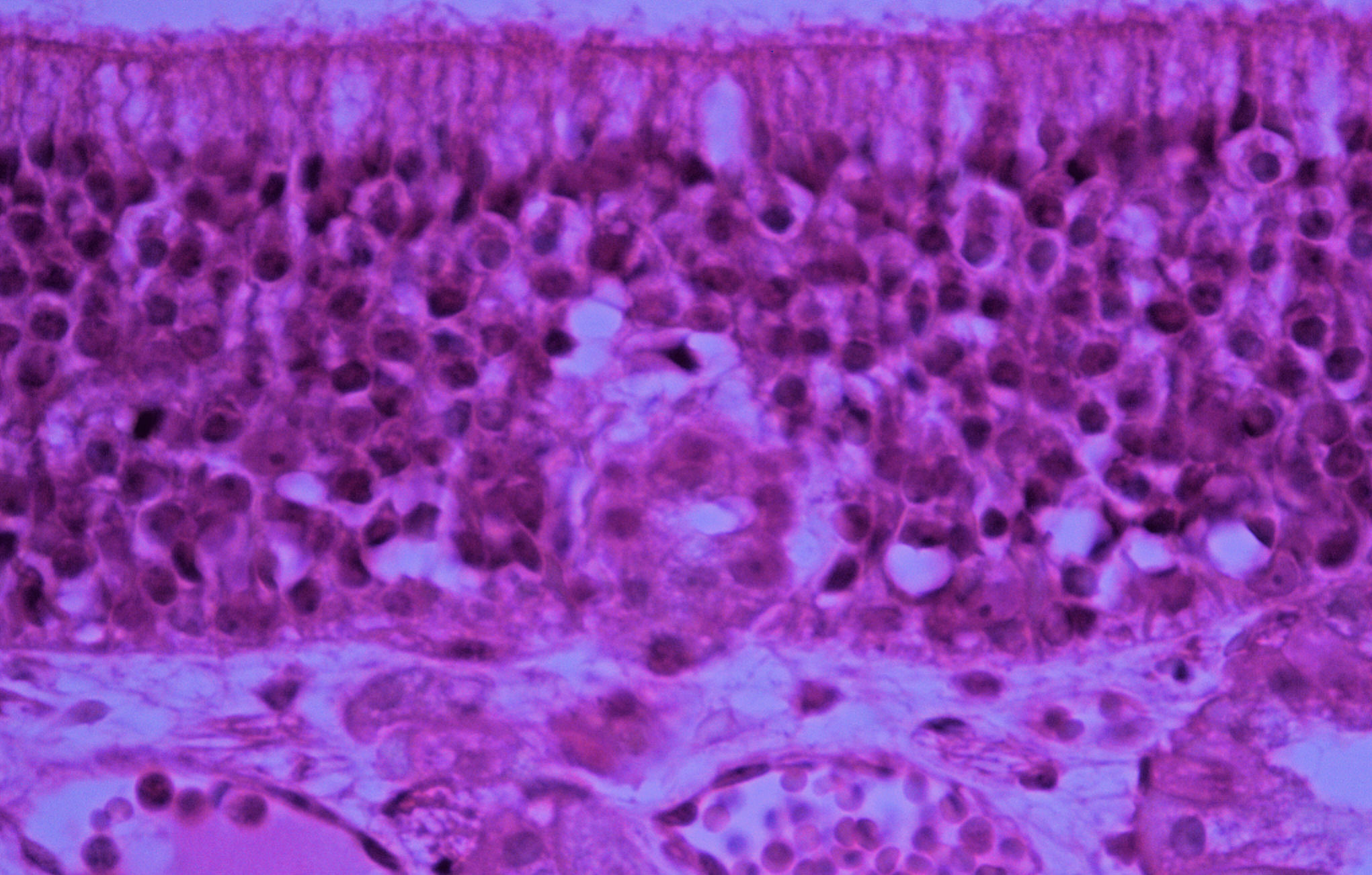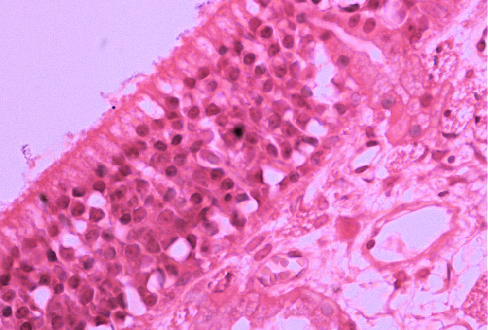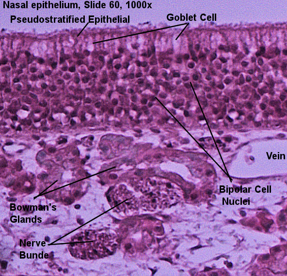
This page has histology of the nasal epithelium. The olfactory nerve extends rostrally from the olfactory cortex (frontal lobe) and ends at the olfactory bulb. In the olfactory bulb, the neurons of the olfactory nerve junction with axons of bipolar neurons which go through tiny foramen in the ethmoid bone and enter the nasal cavity. The bipolar cells extend through the olfactory epithelial and their dendrites essentially dangle into the nasal cavity. The dendrites of the bipolar cells have microvilli (sometimes incorrectly called cilia) which extend the surface area and have different combinations of chemoreceptors on them. The retina has 3 layers of cells which help to convert what you "see" into nural impulses. Immediately adjacent to the black choroid you will see the rod and cone cells. Above the cells you will see the nuclei layer for the rods and cones. This should give you a good indication of how many photo receptors are in the retina. The bipolar cells may be hard to identify but you should be able to see the nuclei of the bipolar cells in a distinct layer. The last layer will be the nuclei of the ganglion cells.
The pictures were taken by me over various semesters. The lab book's picture is on the left and unlabeled picthers, which you can scroll through with the arrows, are on your right.Compare the unlabeled pictures with the labeld picture and try to find the structures from your lab book's picture. Then select one of the pictrues to draw in your lab book.
| Lab Book Image | Student Images |
|---|---|
|
|
|