Supporting Connective Tissue Histology | Histology Home Page | Site Home Page
Compact Bone Conective Tissue
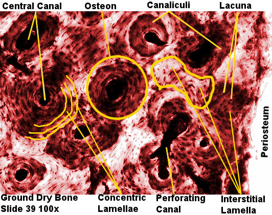
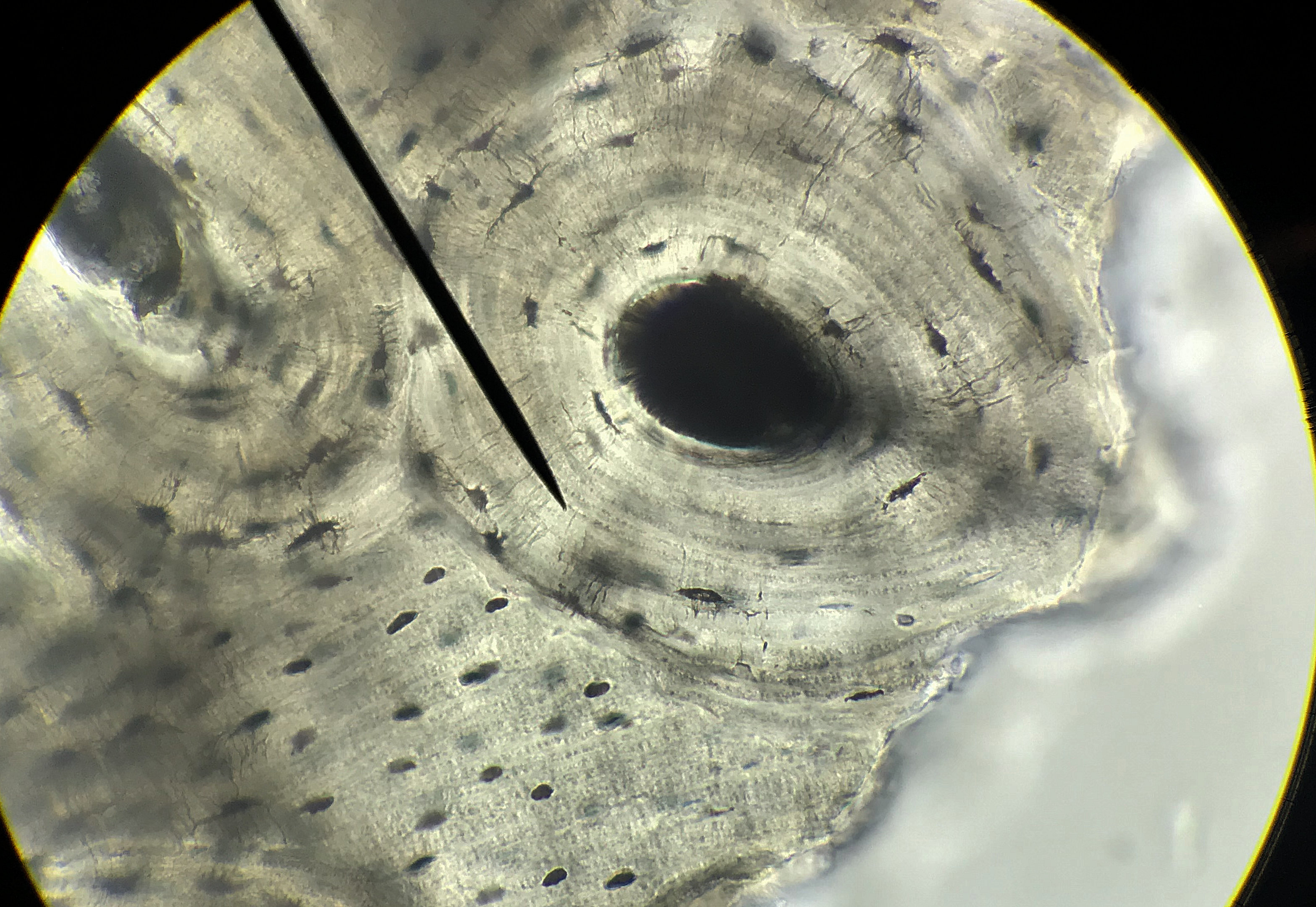
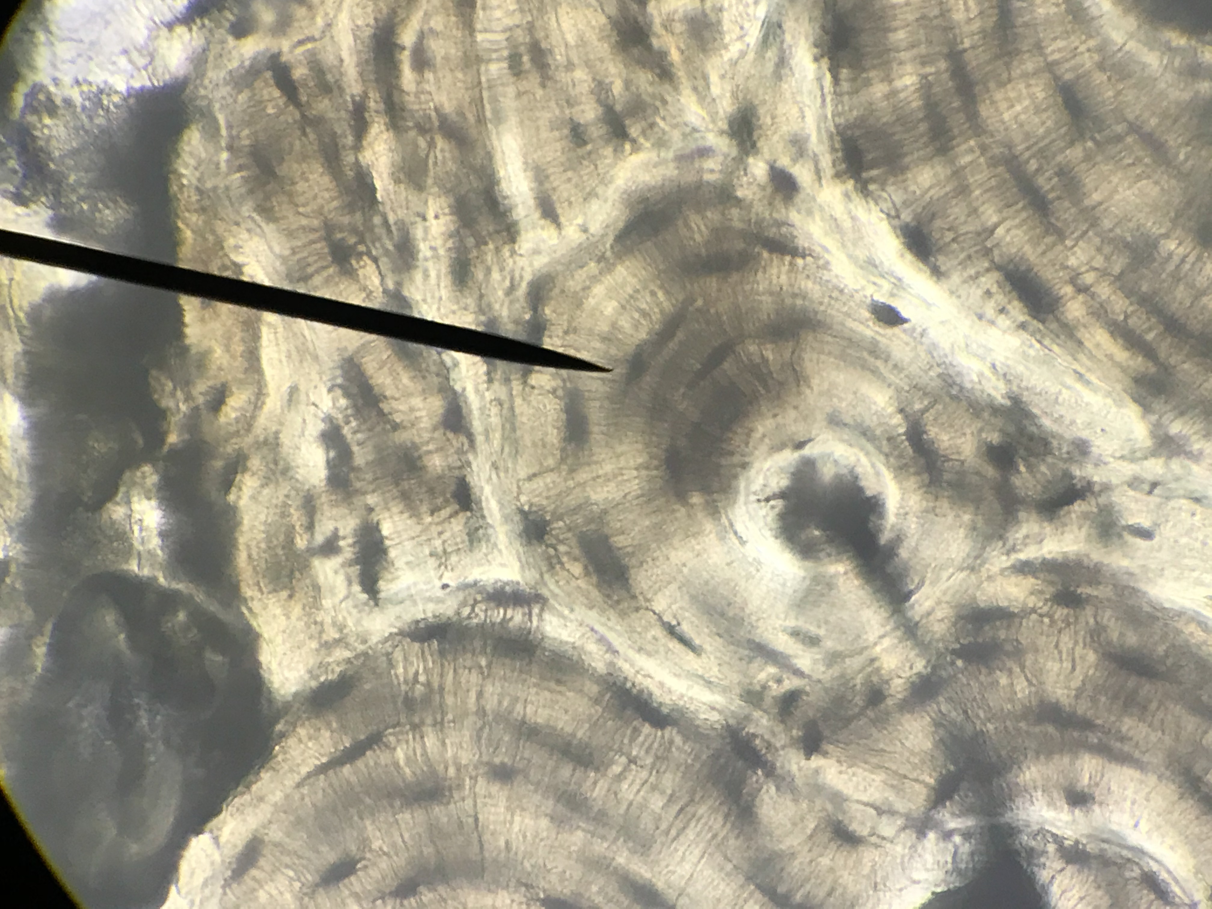
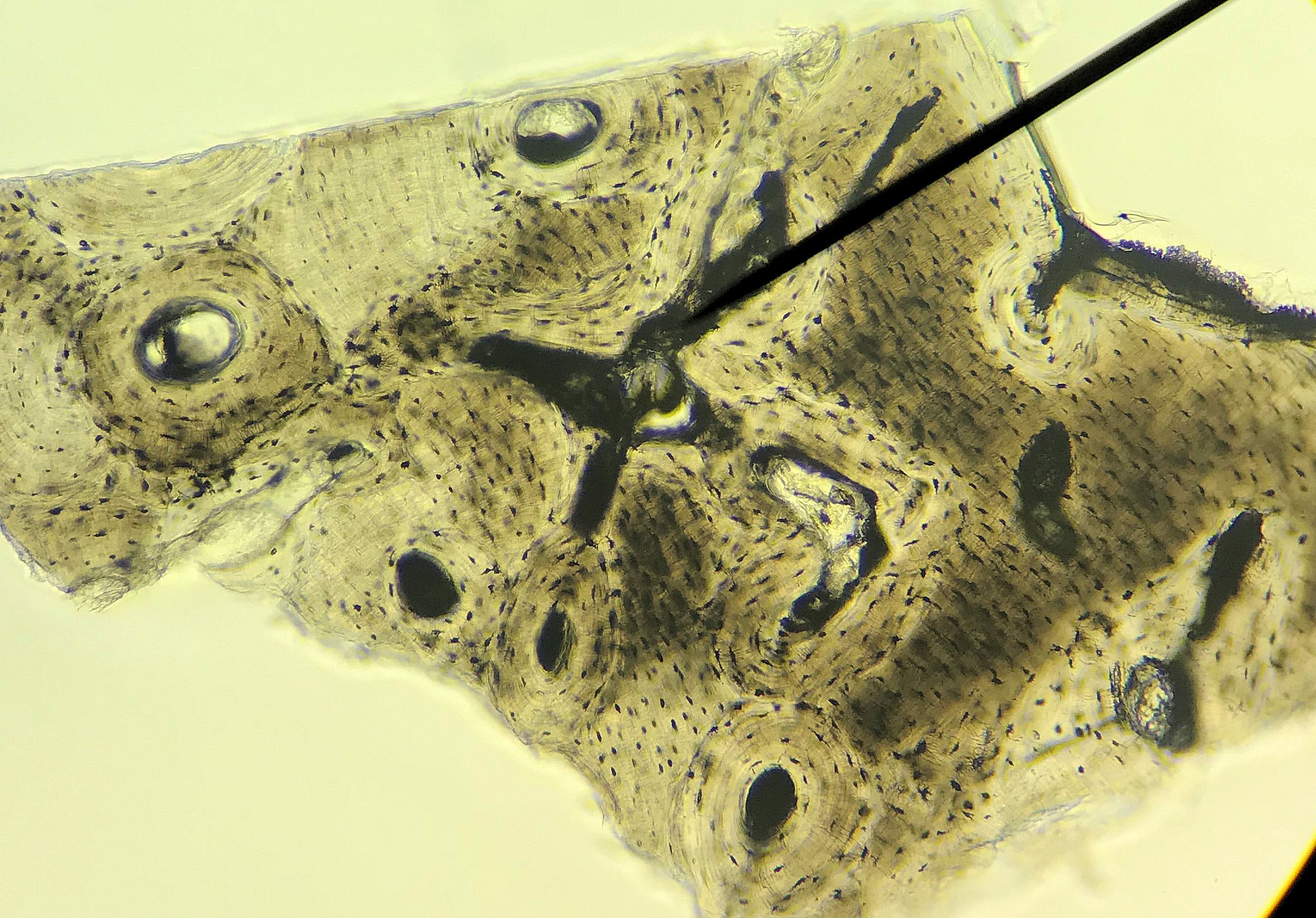
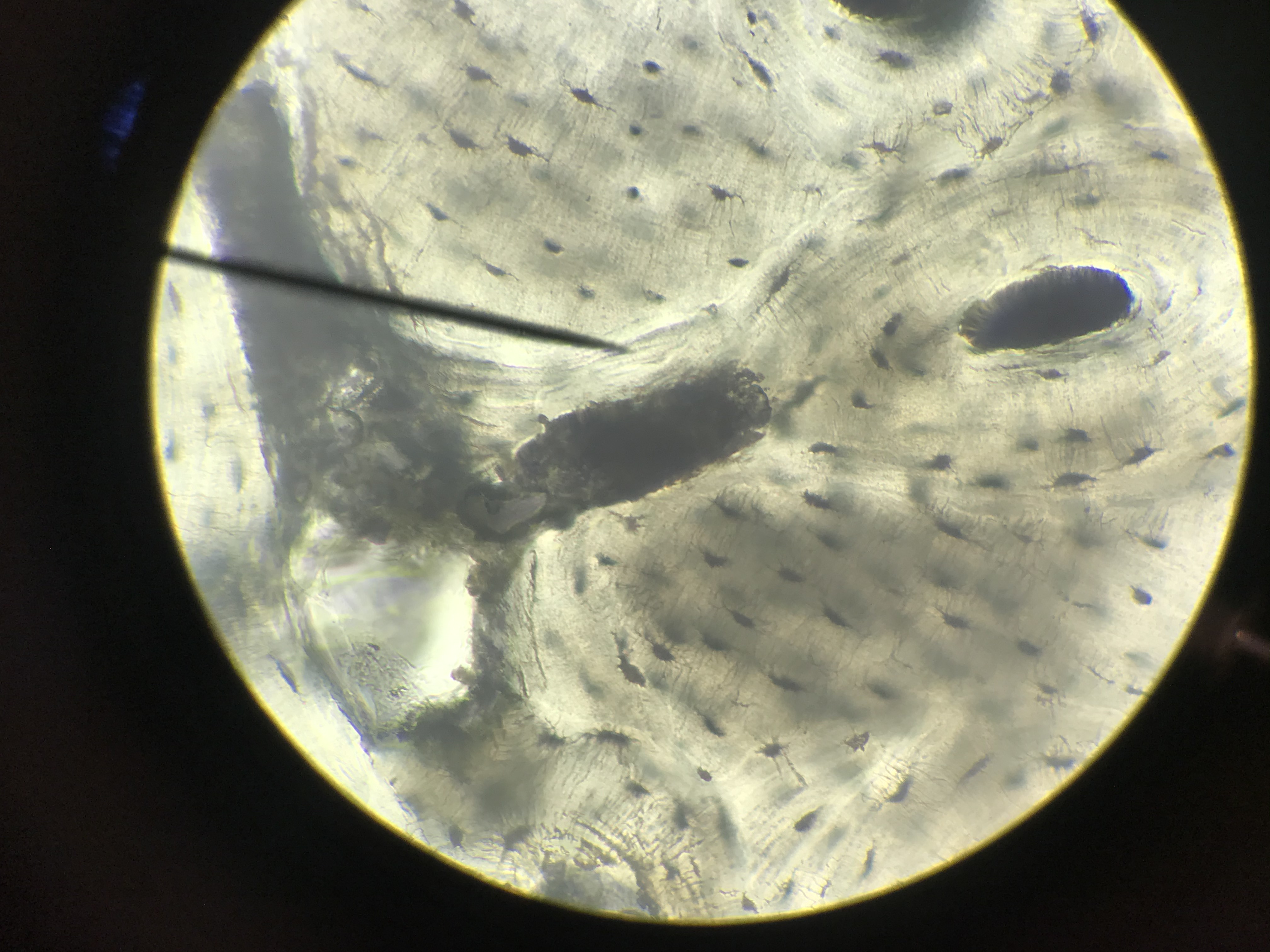
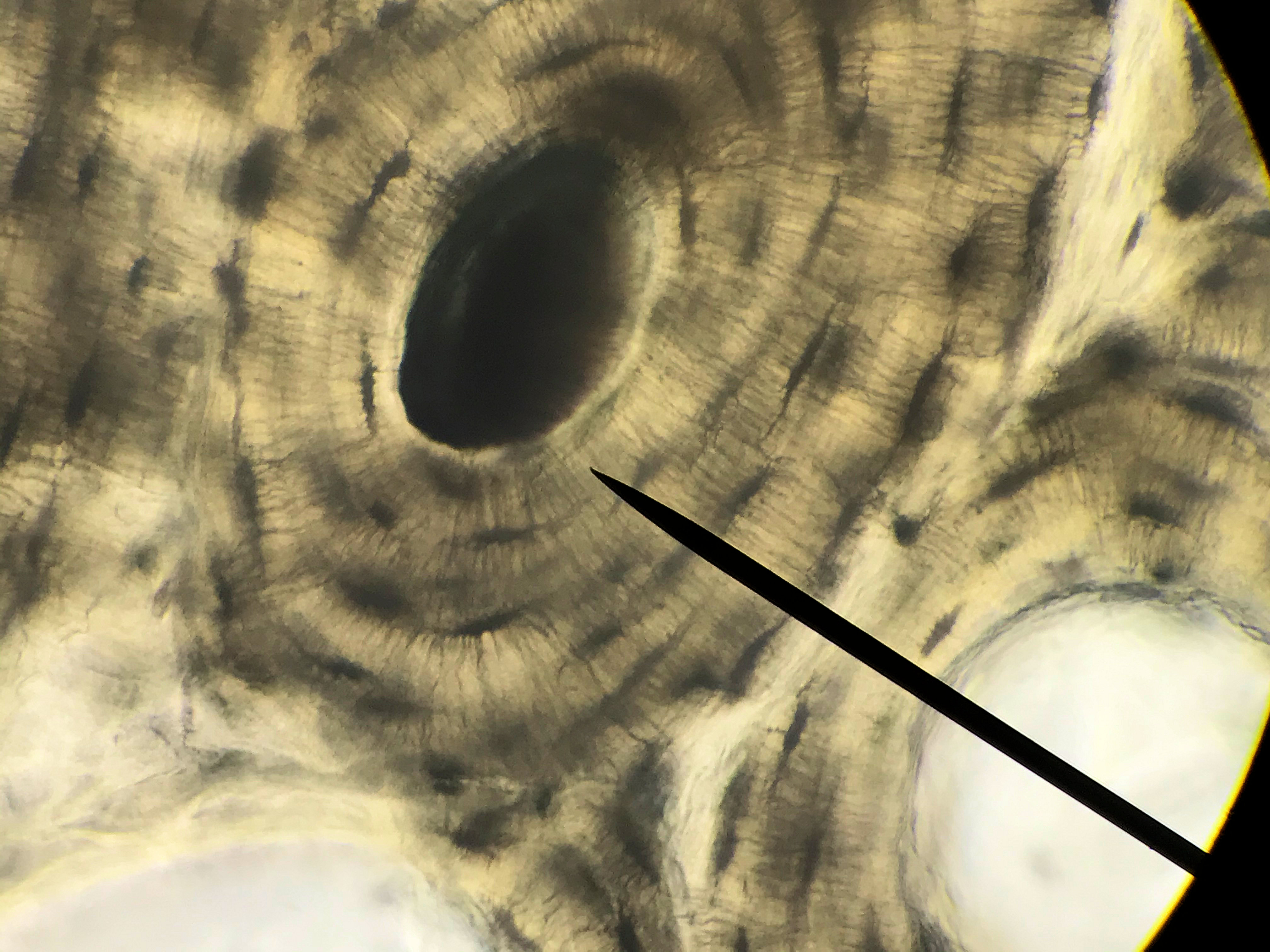
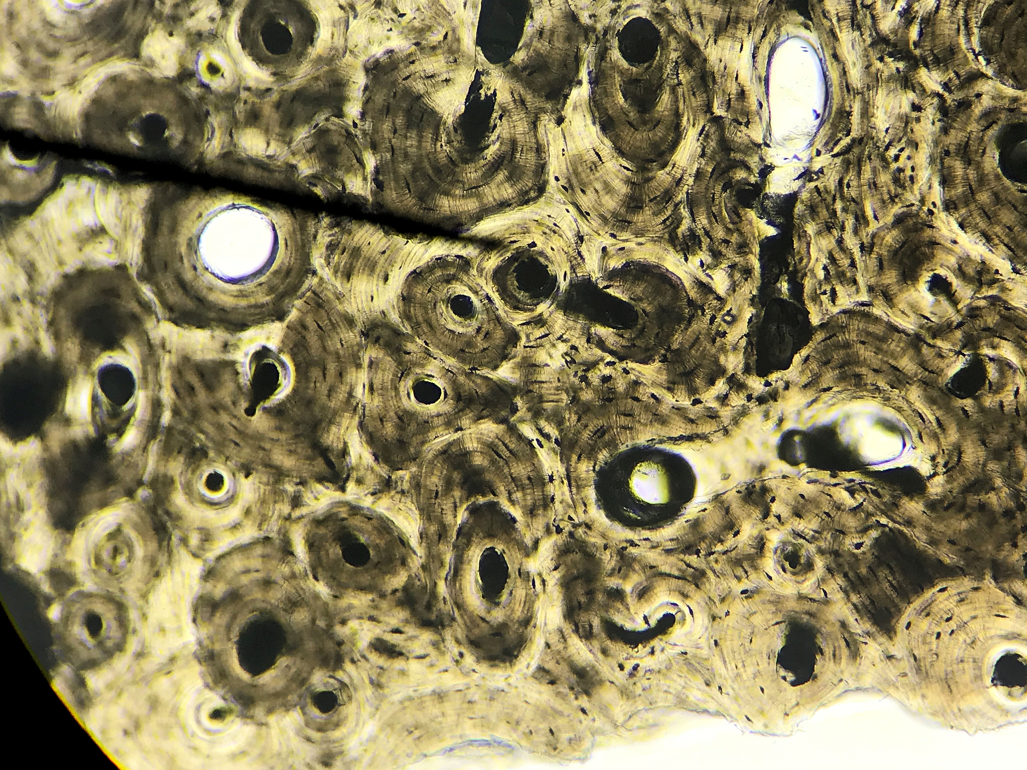
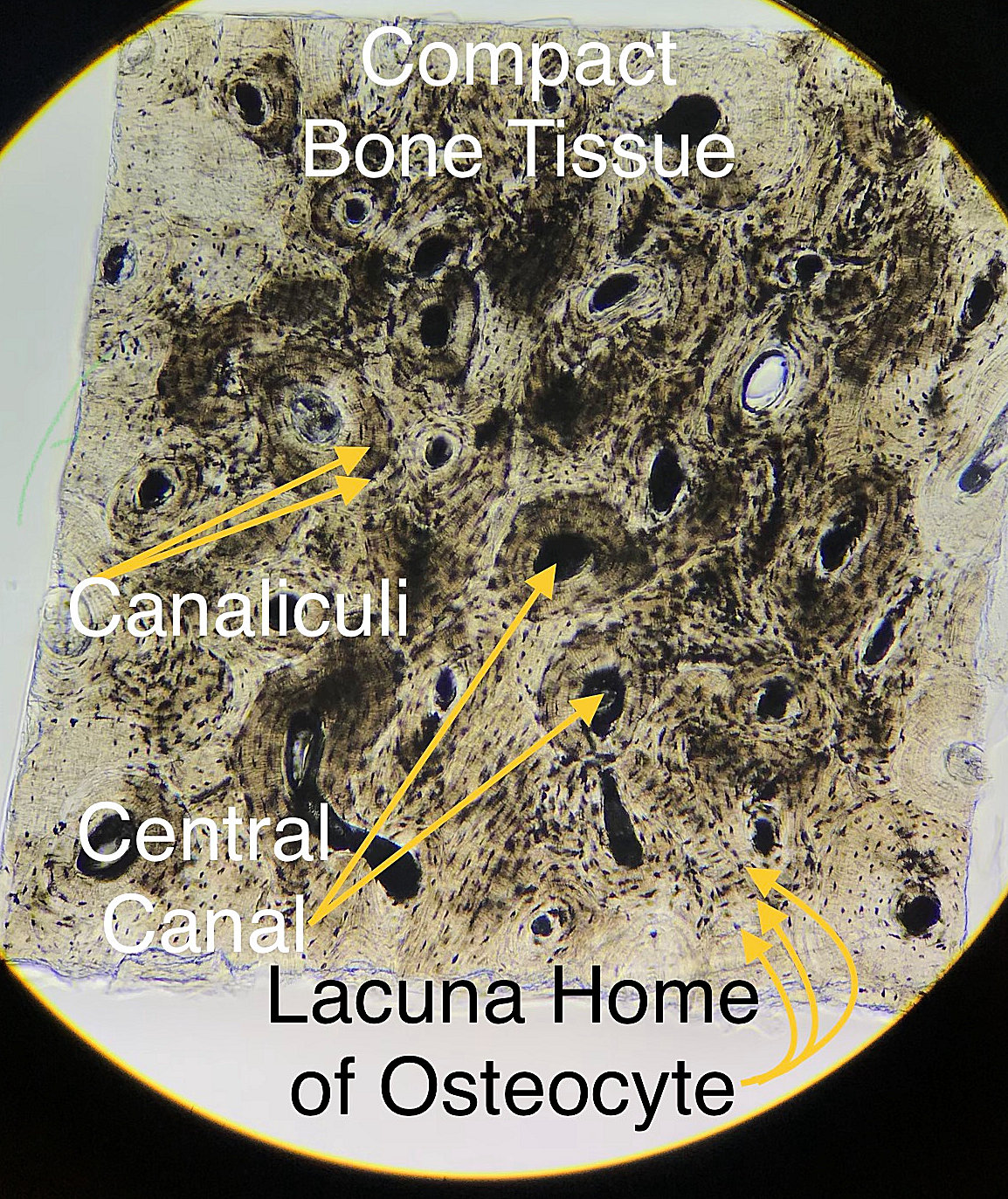
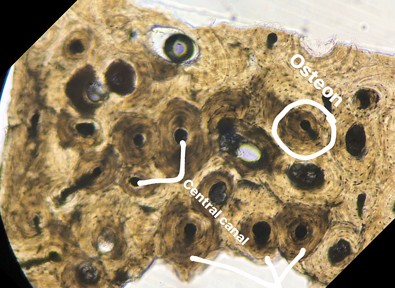
Compact bone gets its name due to its dense compressed structure. Osteocytes are located in lacuna and the lacuna tend to be found in concentric circles around a central (Harversian) canal. The matrix found between the lacuna form concentric circles which are called concentric lamella. Osteocytes are in fact star shaped and they have processes that project through the matrix to other osteocytes through canals called canaliculi. The canaliculi allow for nutrients/oxygen to pass from the central canal to osteocytes in the outer concentric lamella. In compact bone, this organization gets repeated several times to form structures called osteons. Periodically, 2 central canals are linked by a perforating (Volkmann's) canal that allow for blood to flow between the vessels in the central canals. Lastly, bone is constantly being remodeled. Thus, occasionally you will see remnants of old osteons in the form of interstitial lamella.
This model and this model show
what you are supposed to be looking at for this slide.
Slides on this page were made by students between the spring of 2018 and the spring of 2020. Go through
the diffrent student pictures and compare them to your lab book picture. Then slect one to draw on paper. Be sure
to label the cells, lacuna, fibers and other structures.
| Lab Book Image |
Student Images |
|
1 / 8
2 / 8
3/ 8
4 / 8
5 / 8
6 / 8
7 / 8
8 / 8
❮
❯
|

