Supporting Connective Tissue Histology | Histology Home Page | Site Home Page
Fibrocartilage Conective Tissue
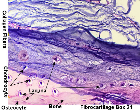
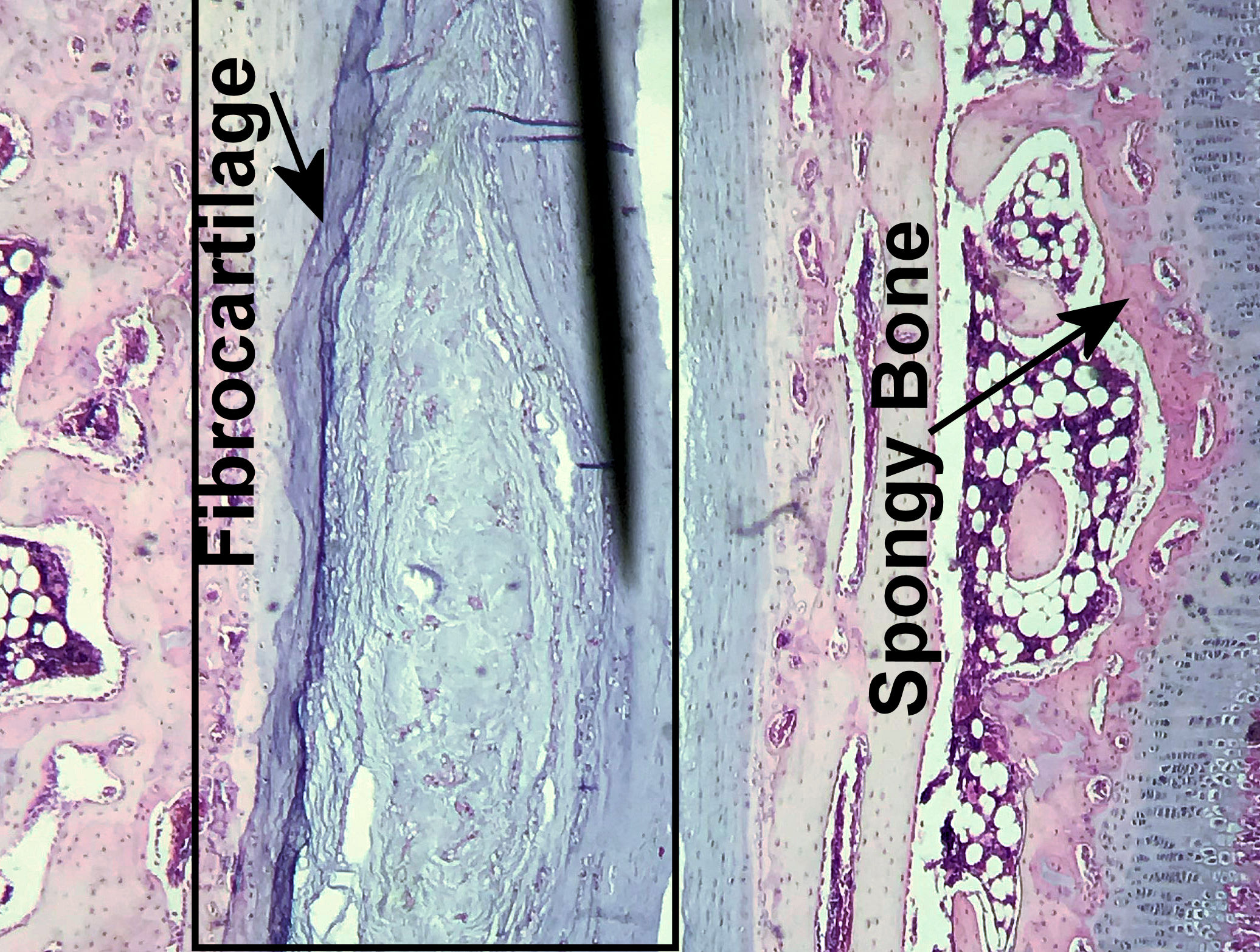
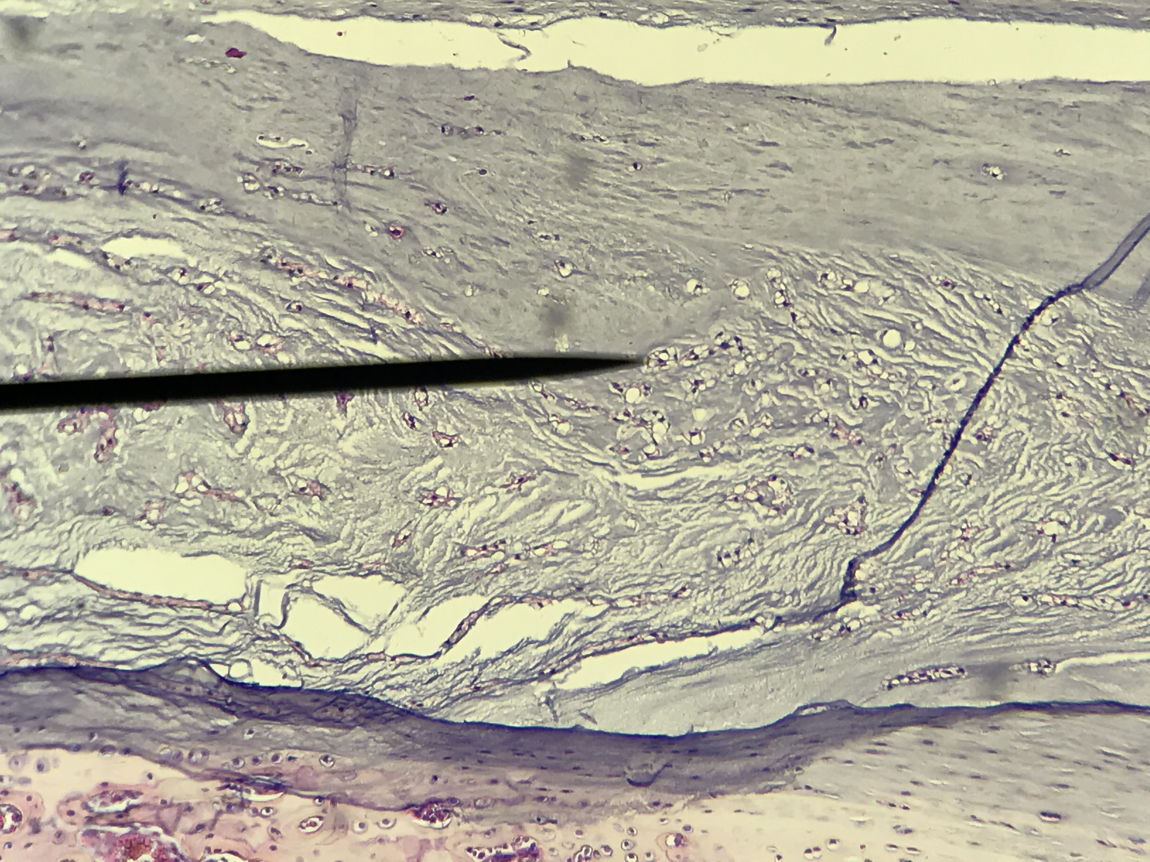
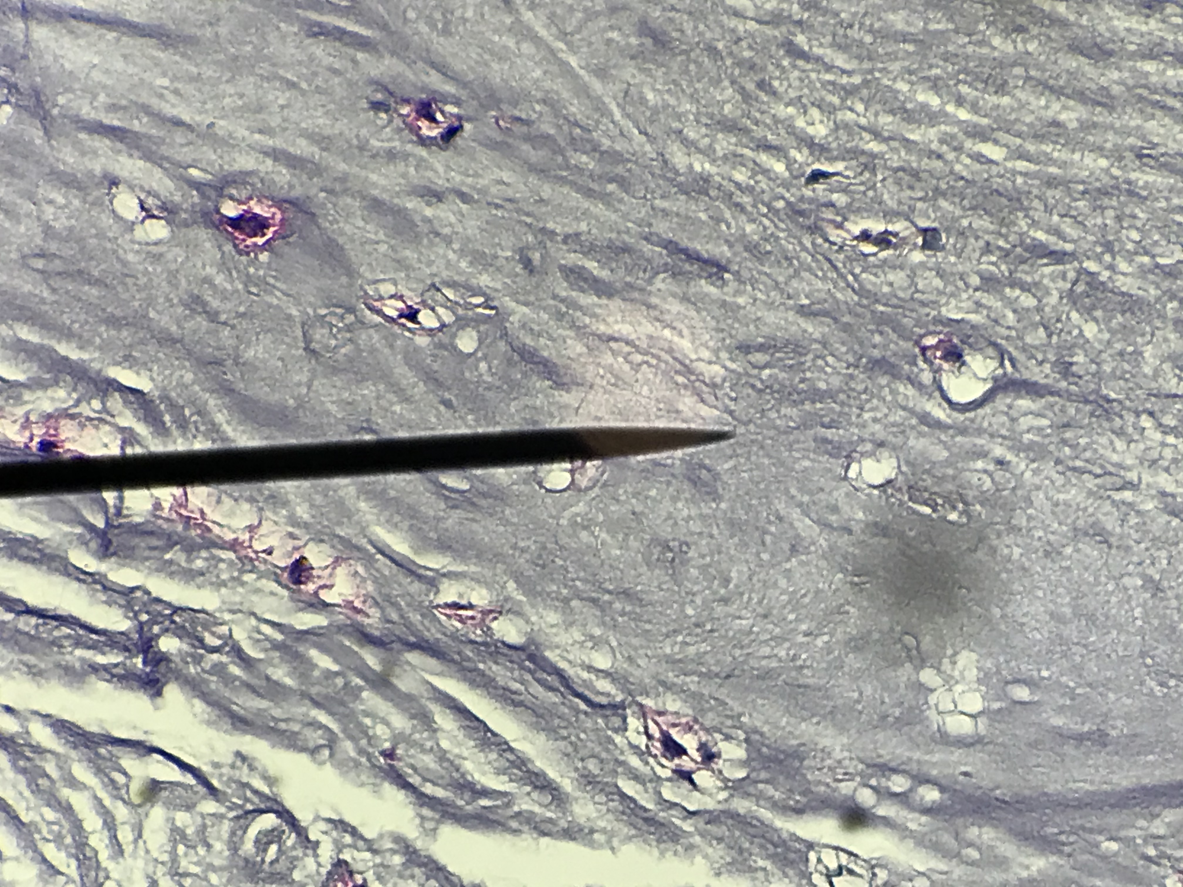
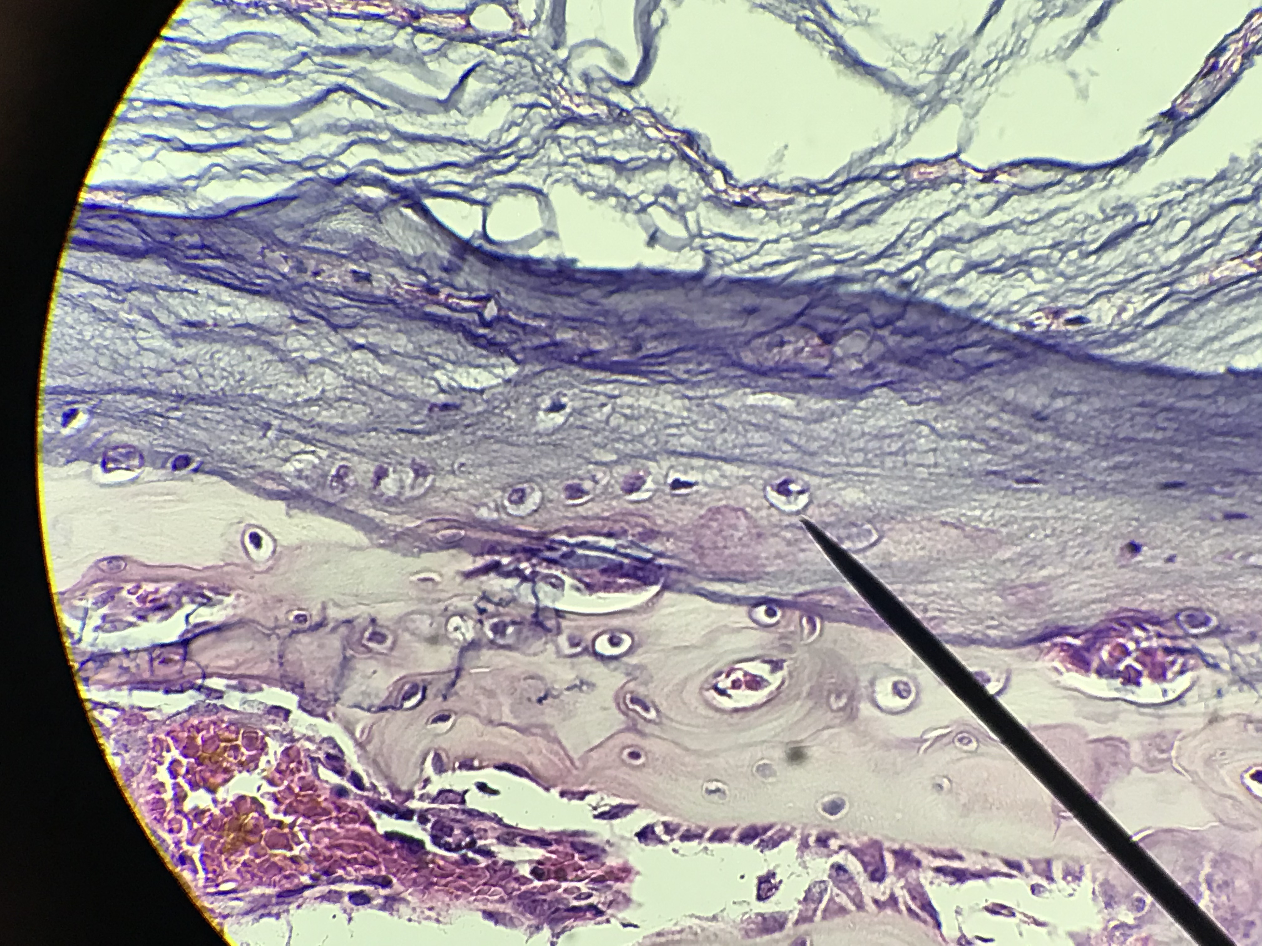
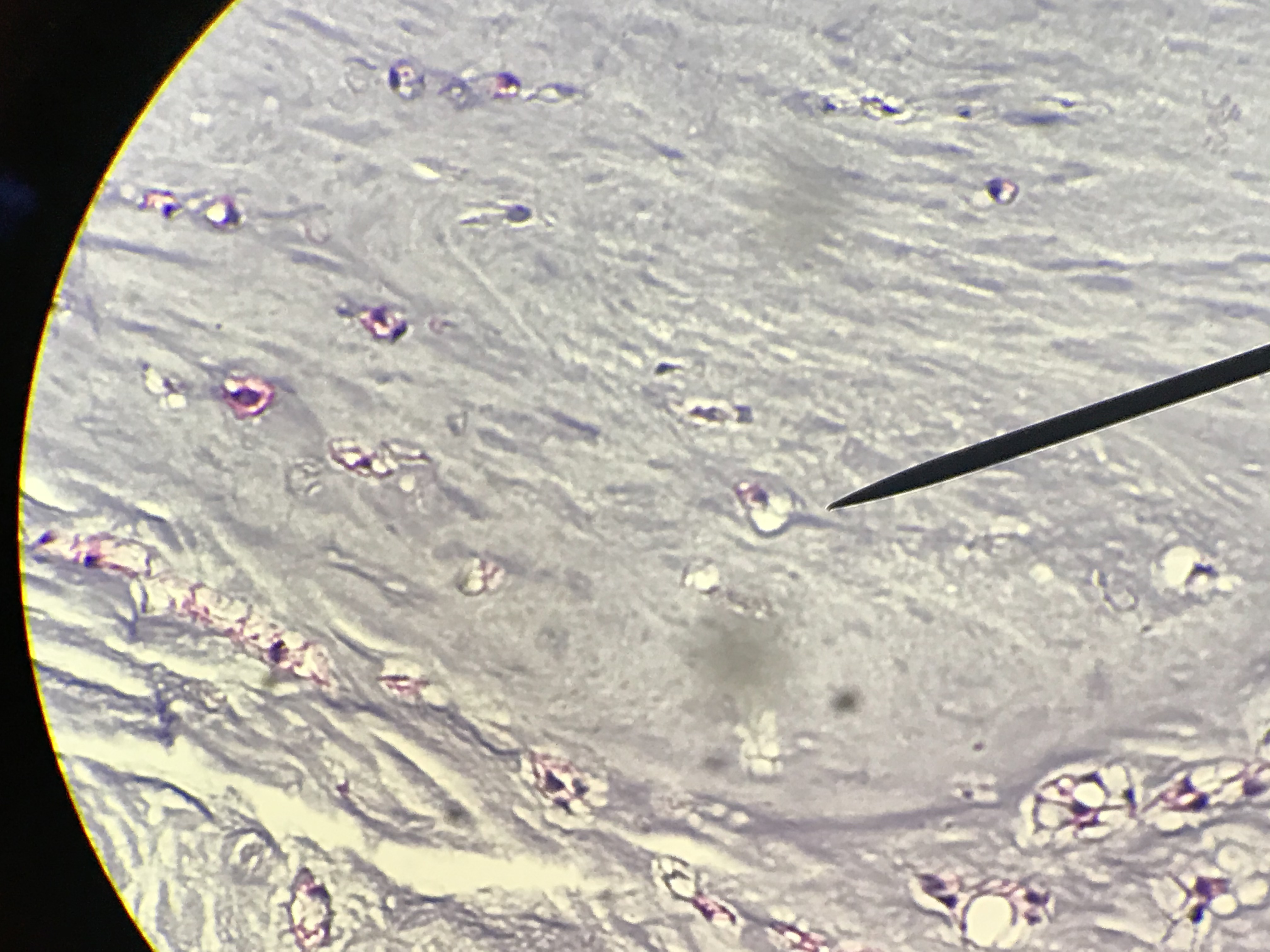
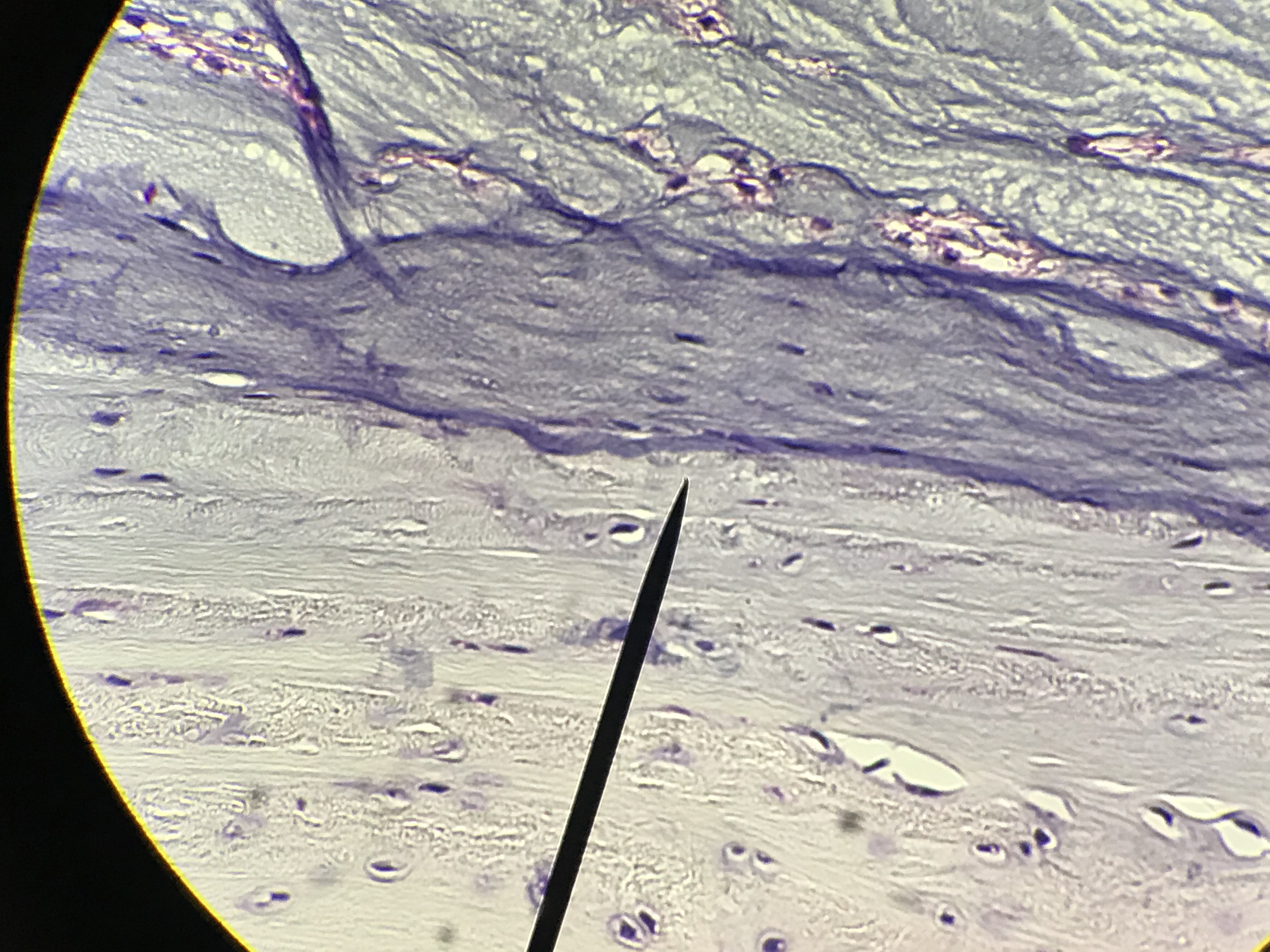
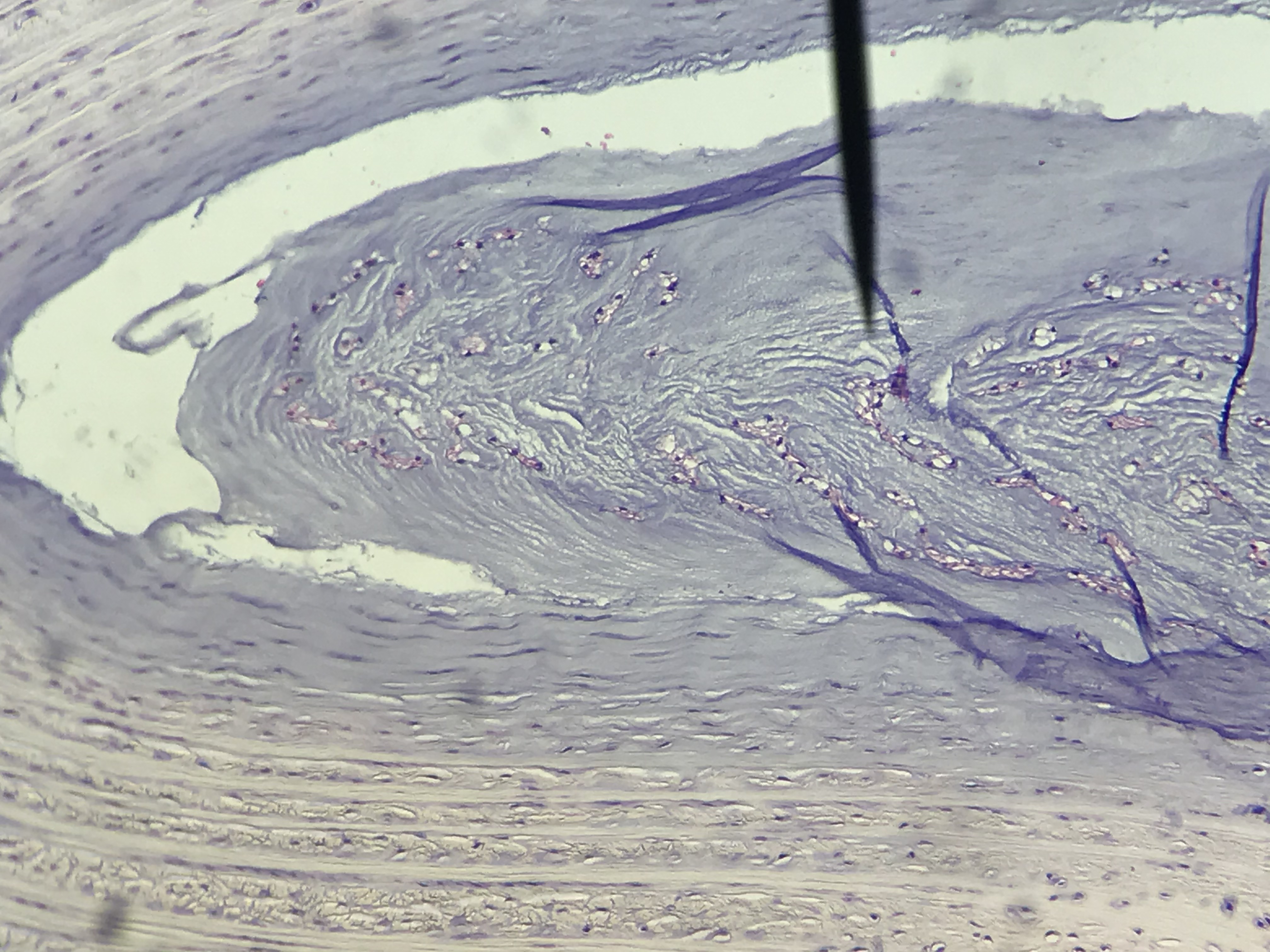
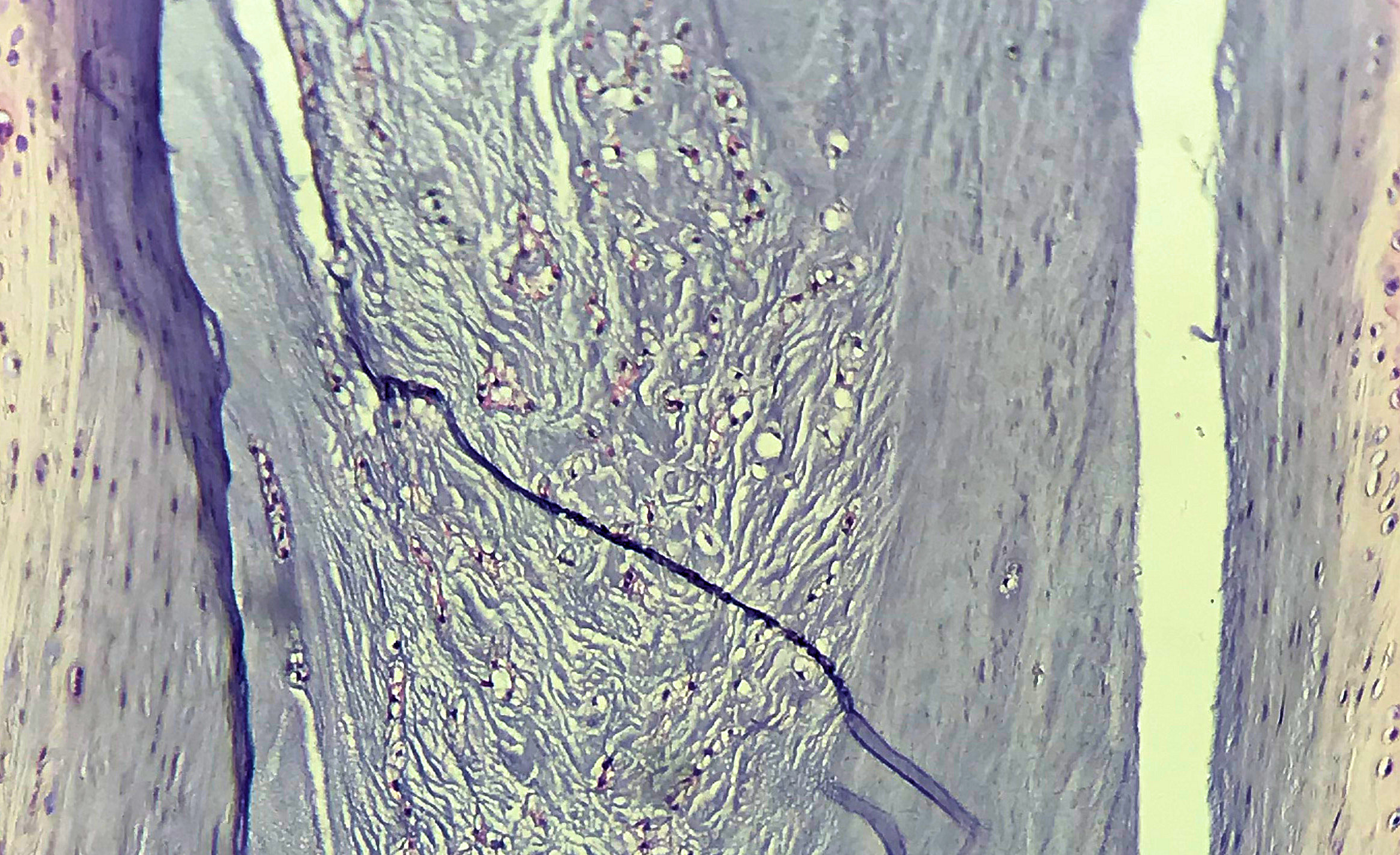
. Fibrocartilage has white (type I) collagen fibers in addition to the type II collage fibers in its matrix. These type I fibers are stain visibly and resemble dense regular connective tissue. The fibers provide more support to the matrix and resistance to compression. This cartilage is observed in the pubic symphysis and the intervertebral disks. It is also found in joints that contain articular discs such as the knee. Unfortunately on histological slides, this Fibrocartilage blends with surrounding tissues.
Slides on this page were made by students between the spring of 2018 and the spring of 2020. Go through
the diffrent student pictures and compare them to your lab book picture. Then slect one to draw on paper. Be sure
to label the cells, lacuna, fibers and other structures.
| Lab Book Image |
Student Images |
|
1 / 8
2 / 8
3/ 8
4 / 8
5 / 8
6 / 8
7 / 8
8 / 8
❮
❯
|