Supporting Connective Tissue Histology | Histology Home Page | Site Home Page
Growth Plate
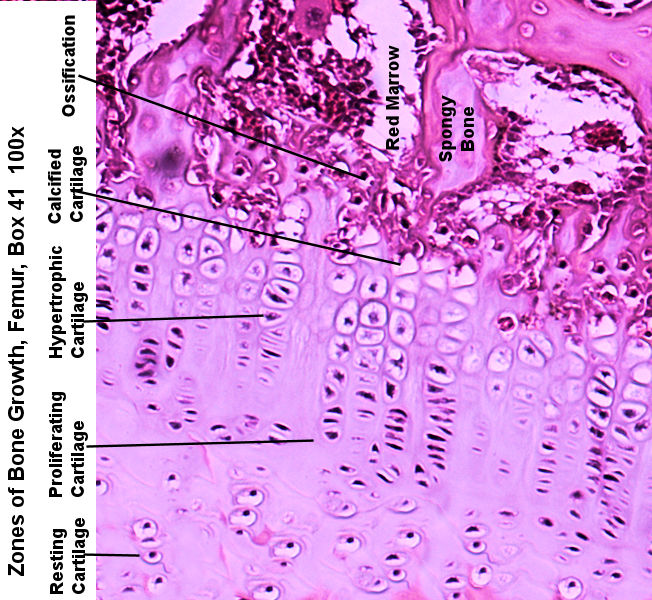
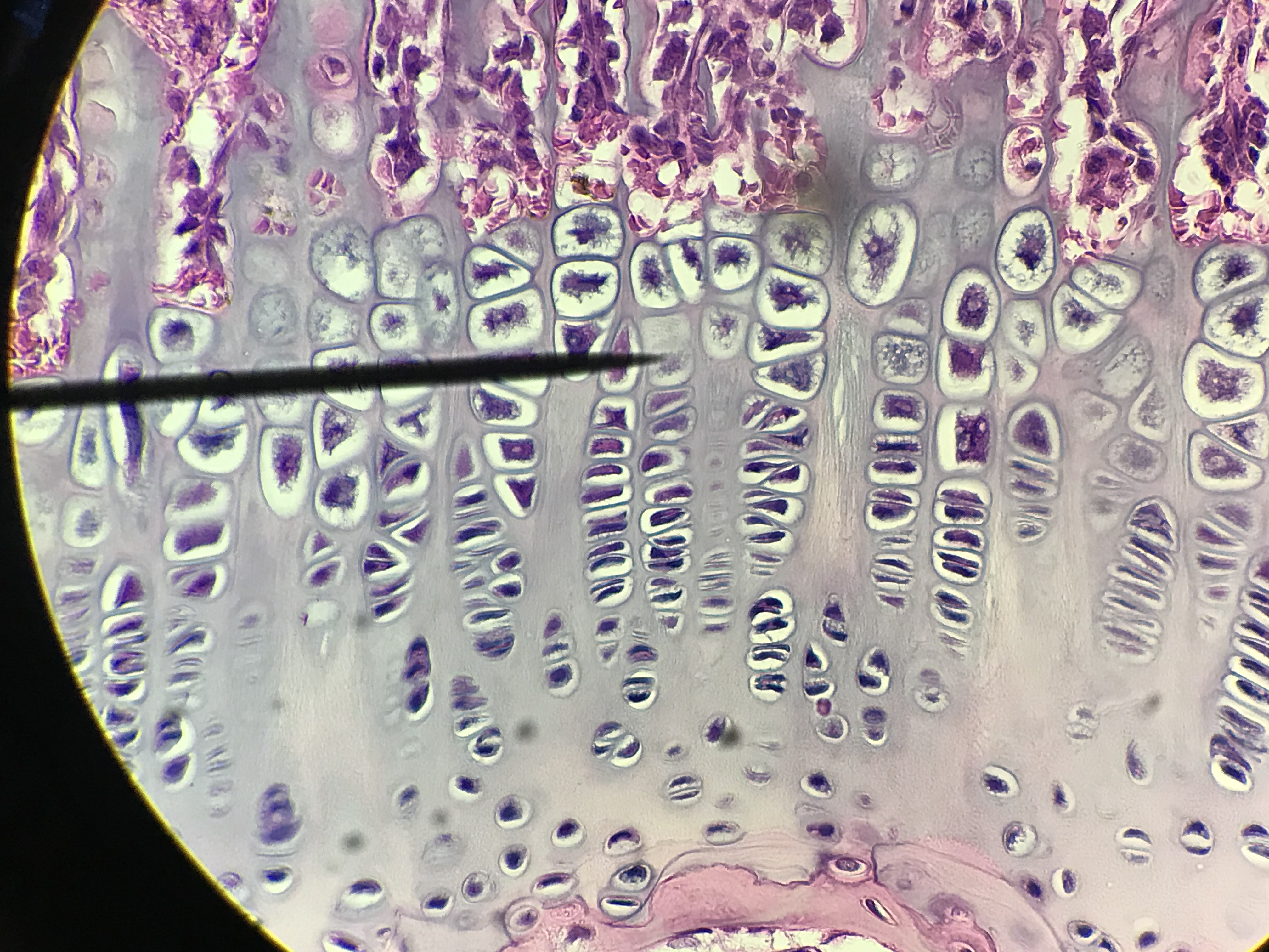
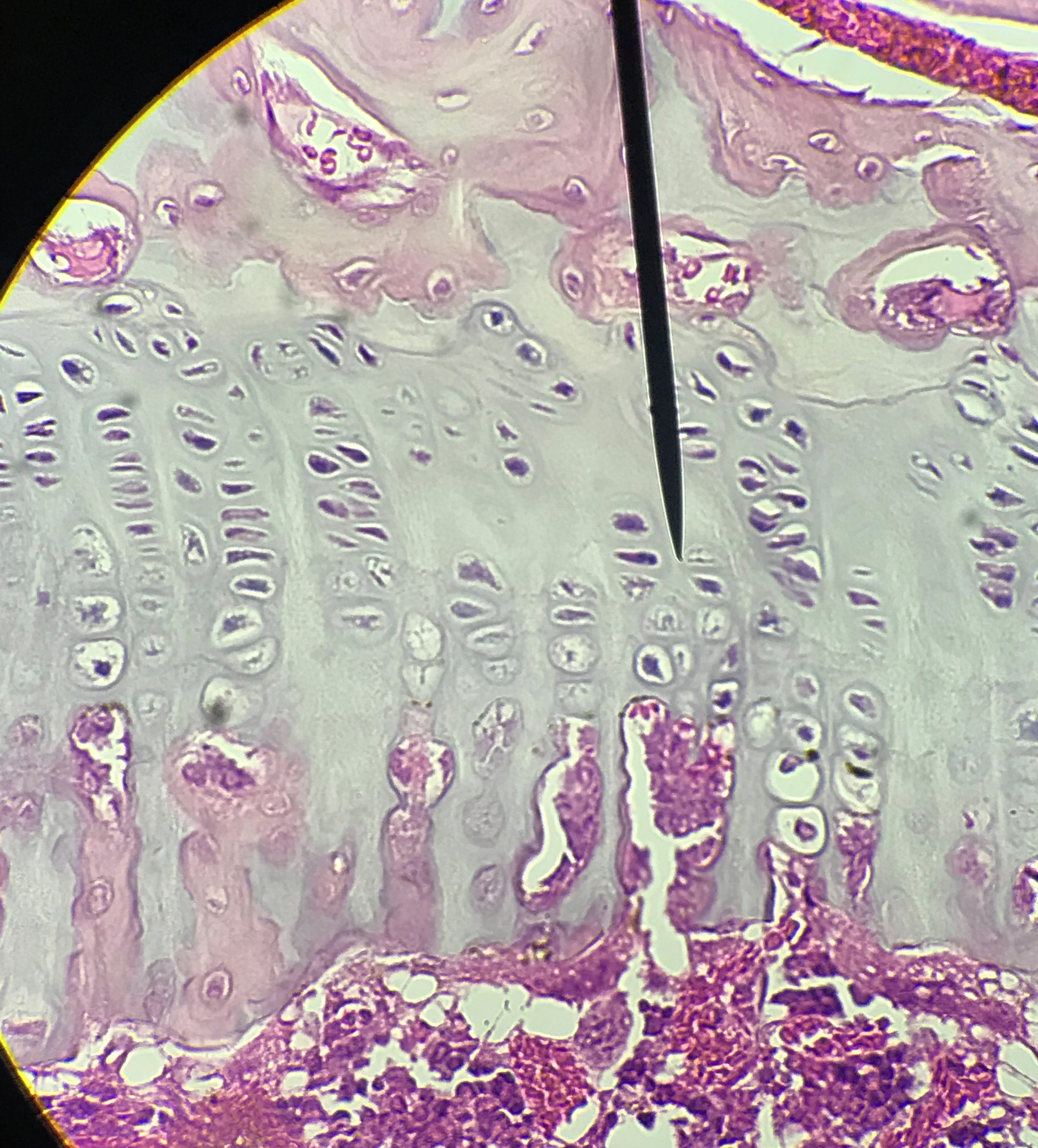
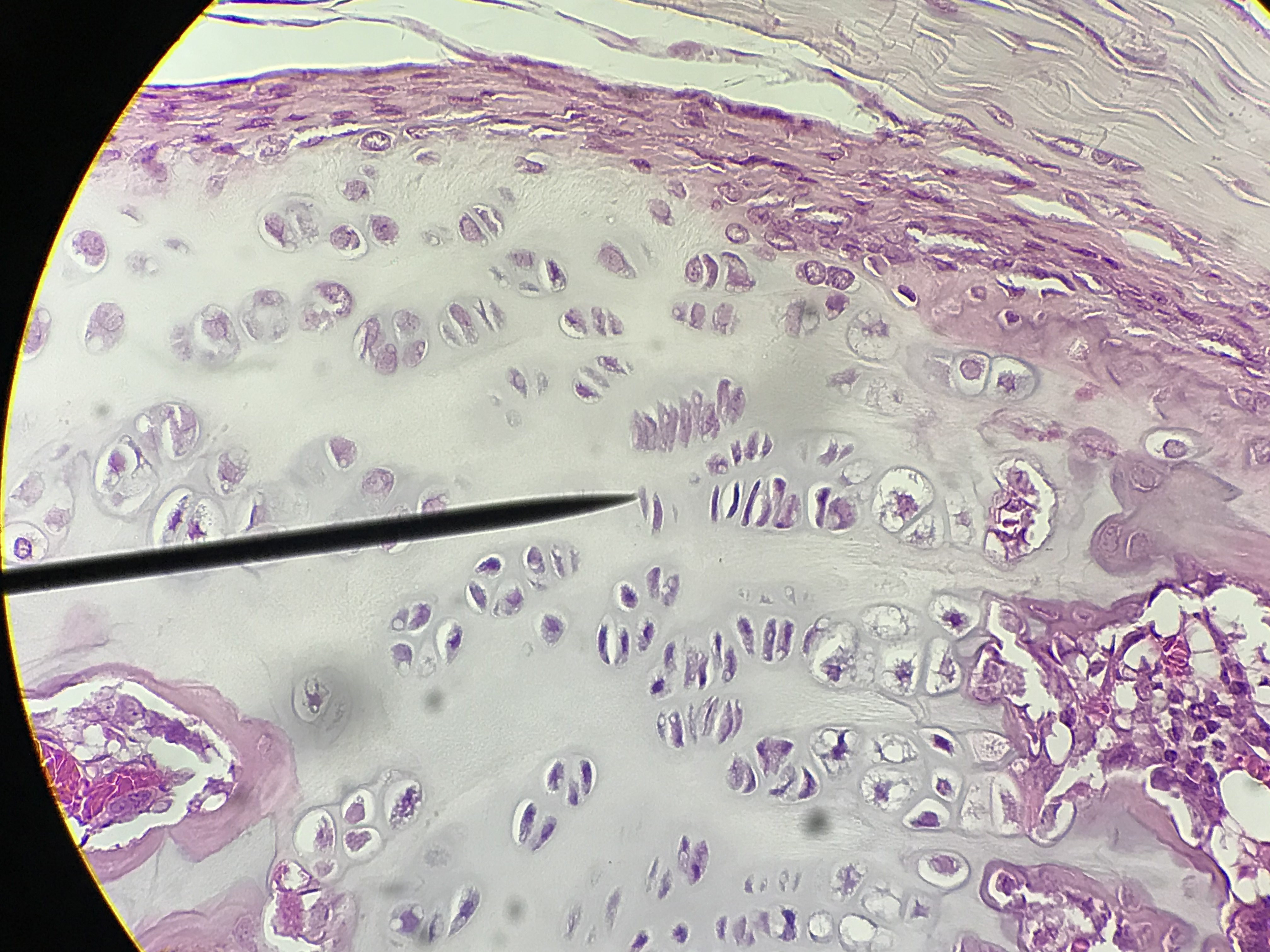
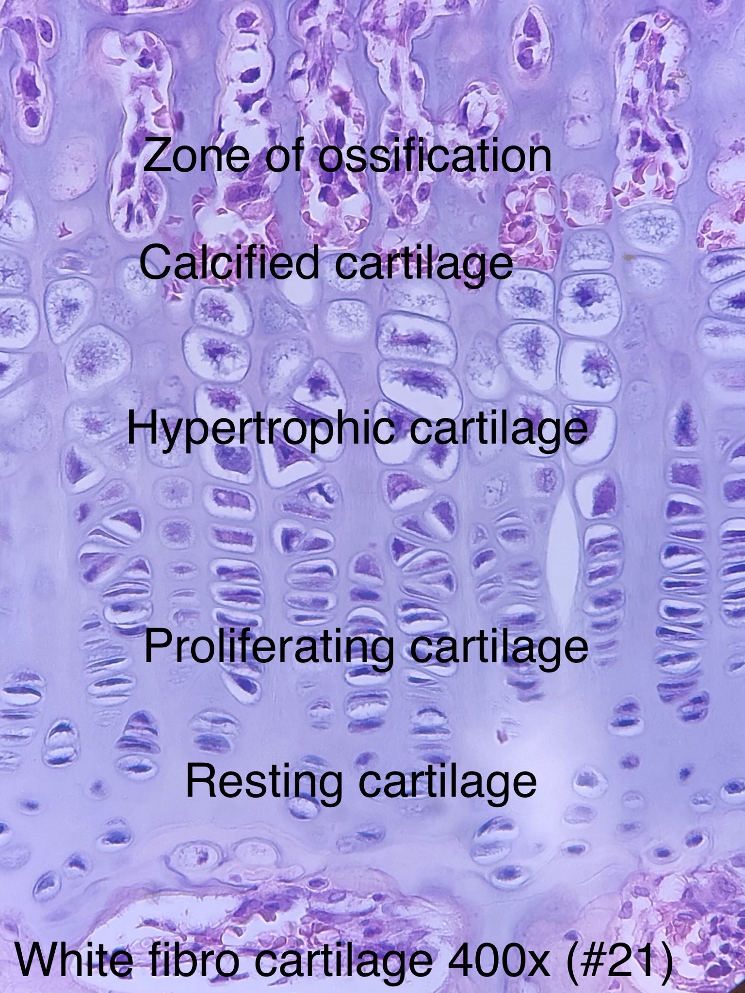
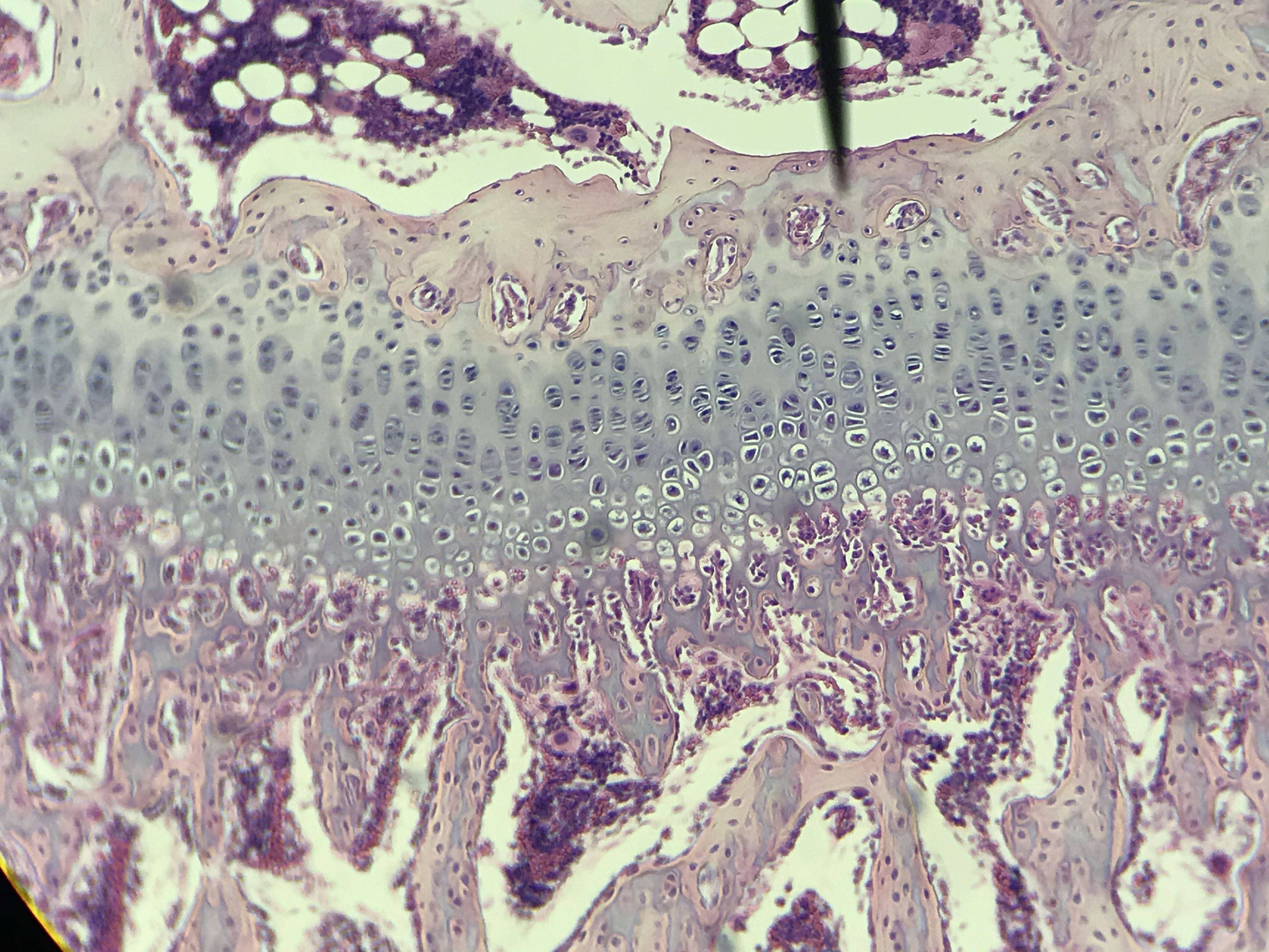
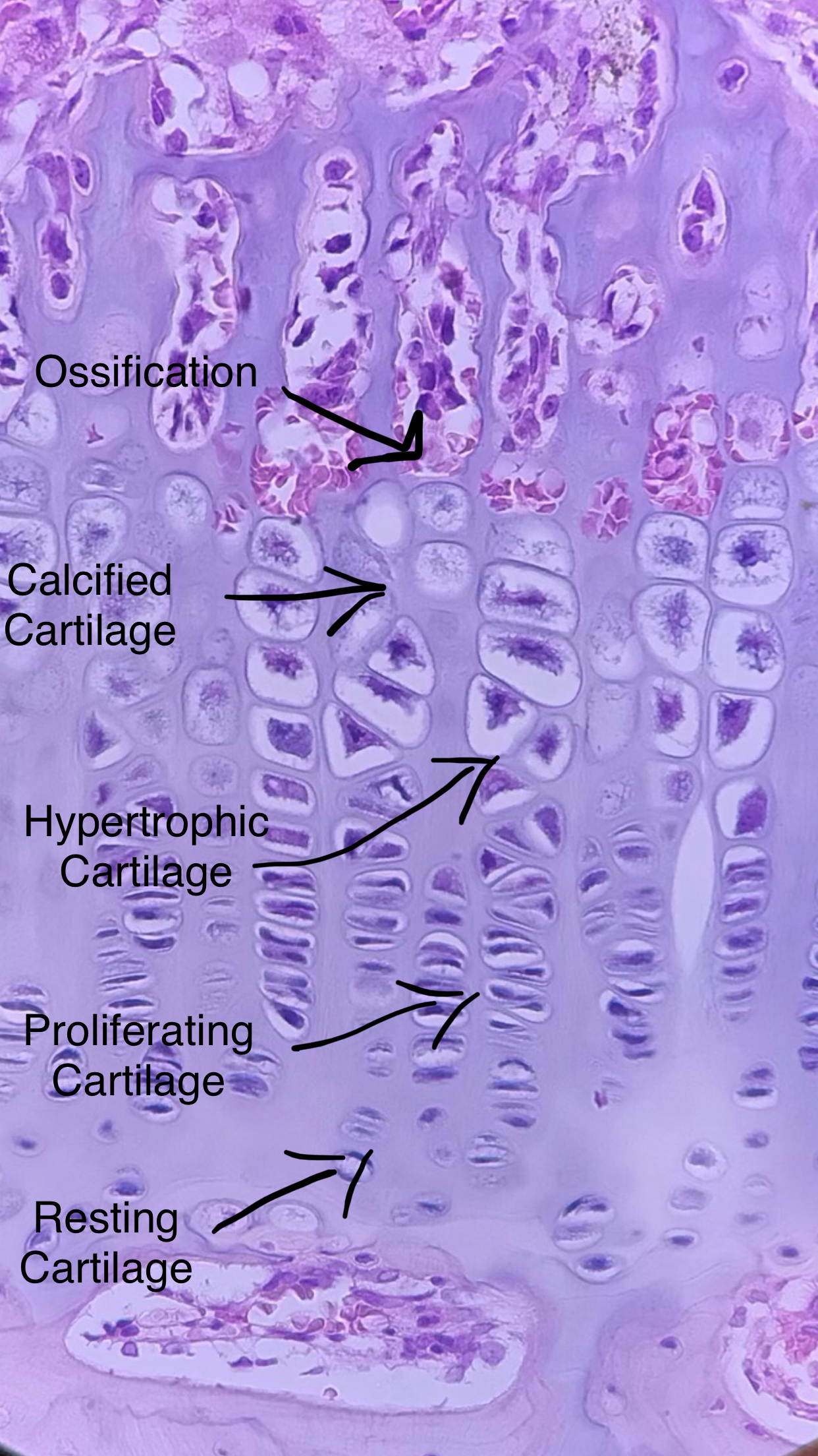
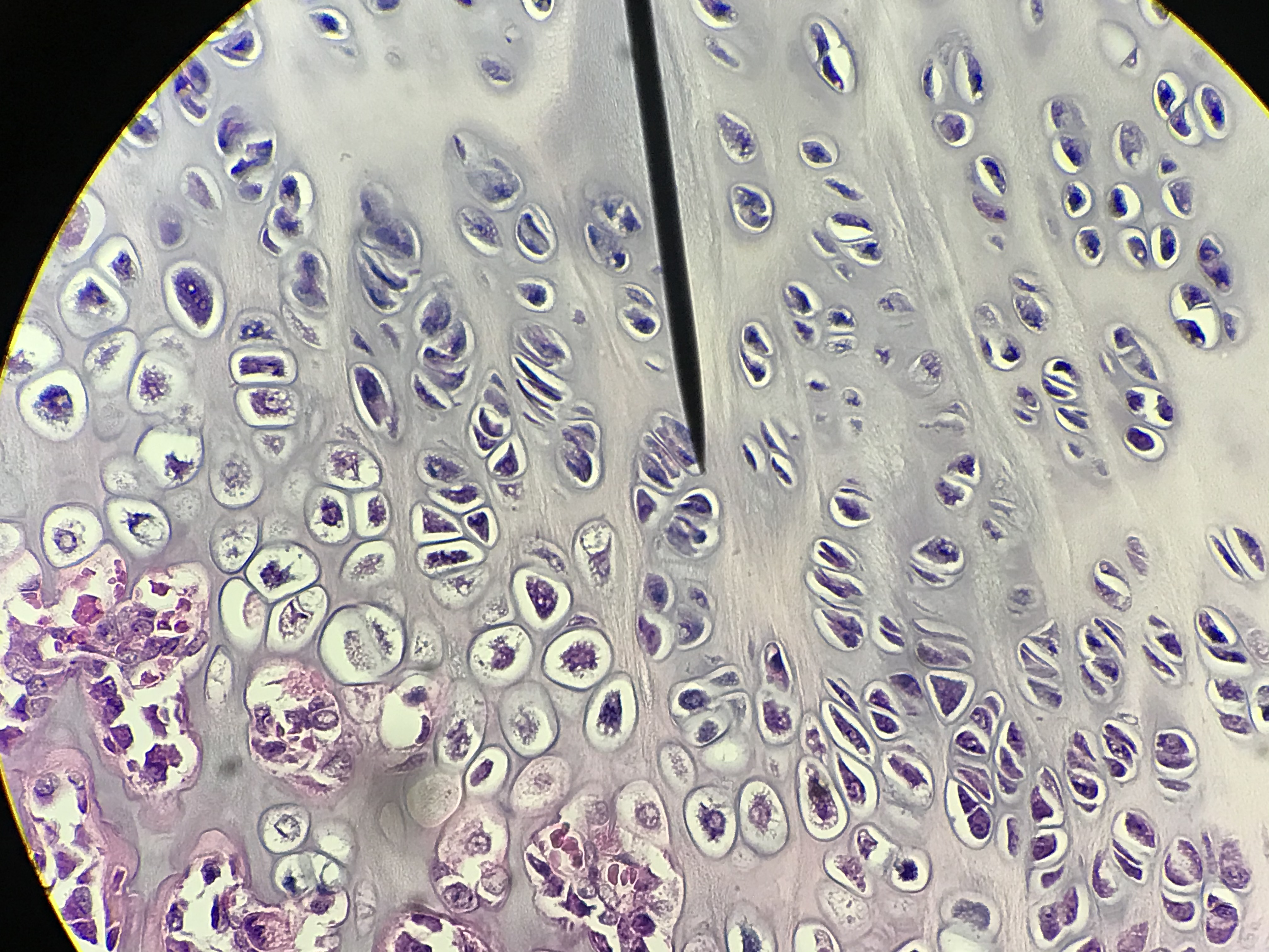
There are typically 5 zones of are visible in the growth plate. The first zone is the zone of resting cartilage and it looks like hyaline cartilage (since it is). When these cells become active, the chondrocytes (cartilage cells) begin rapid mitosis where they line up in neat rows. These rows look like someone stacked piles of pennies on top of each other. The second zone is called the zone of proliferating cartilage. The cells then begin to grow in size. Thus, the next zone is called the zone of hypertrophic cartilage. In zone 3, cell division has stopped and the cells have started to grow in size. Zones 2 and 3 help to elongate the cartilage. In zone 4, the zone of calcified cartilage, the matrix of the cartilage begins to calcify and the chondrocytes die. Therefore, you should observe empty looking lacuna. Last is the zone of ossification. Osteoprogenitor cells, which will become osteoblasts, start to lay down spongy bone and blood vessels
Slides on this page were made by students between the spring of 2018 and the spring of 2020. Go through
the diffrent student pictures and compare them to your lab book picture. Then slect one to draw on paper. Be sure
to label the cells, lacuna, fibers and other structures.
| Lab Book Image |
Student Images |
|
1 / 8

2 / 8
3/ 8
4 / 8
5 / 8
6 / 8
7 / 8
8 / 8
❮
❯
|