Supporting Connective Tissue Histology | Histology Home Page | Site Home Page
Hyaline Cartilage Conective Tissue
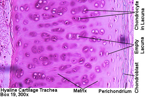
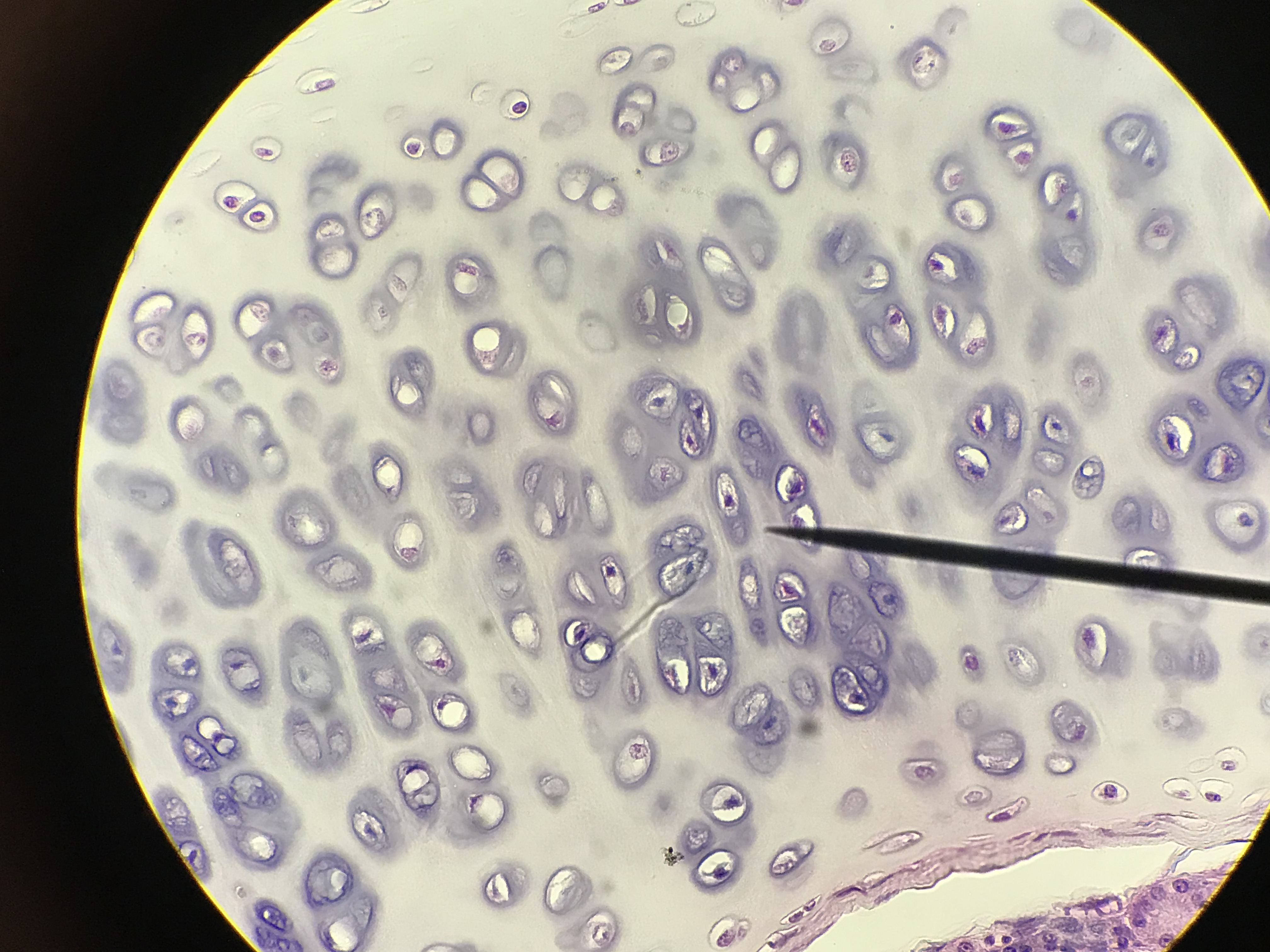
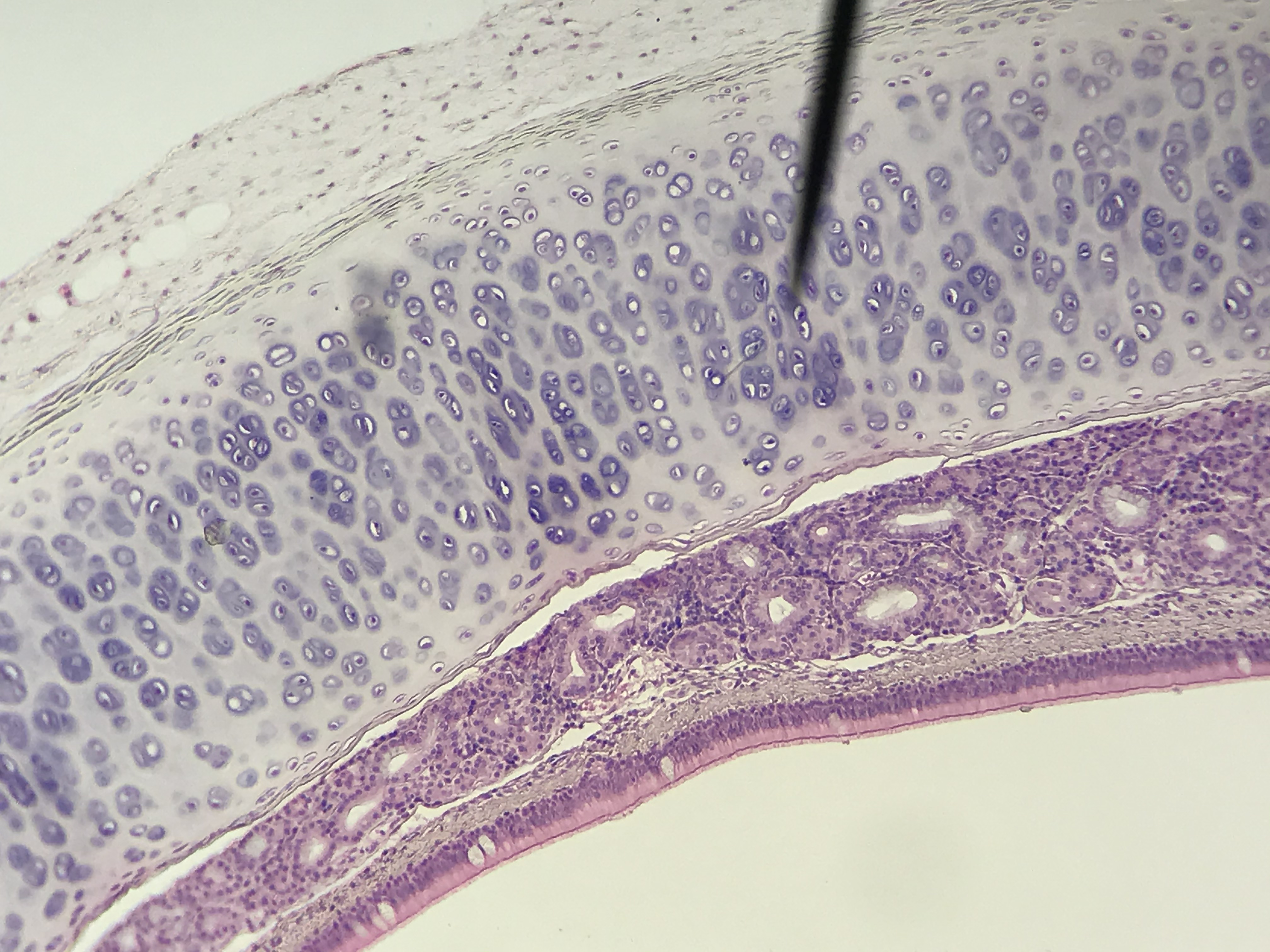
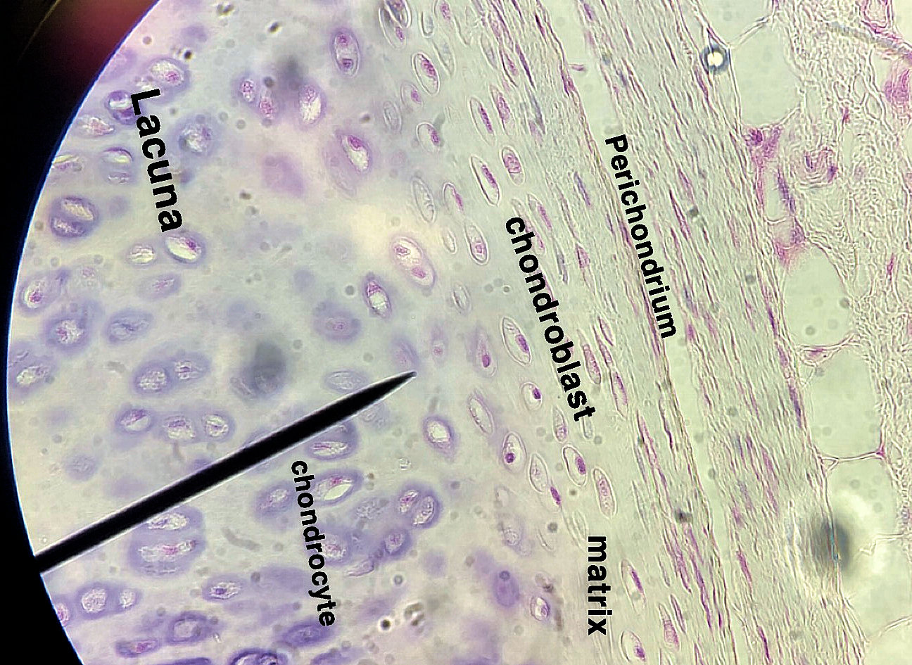
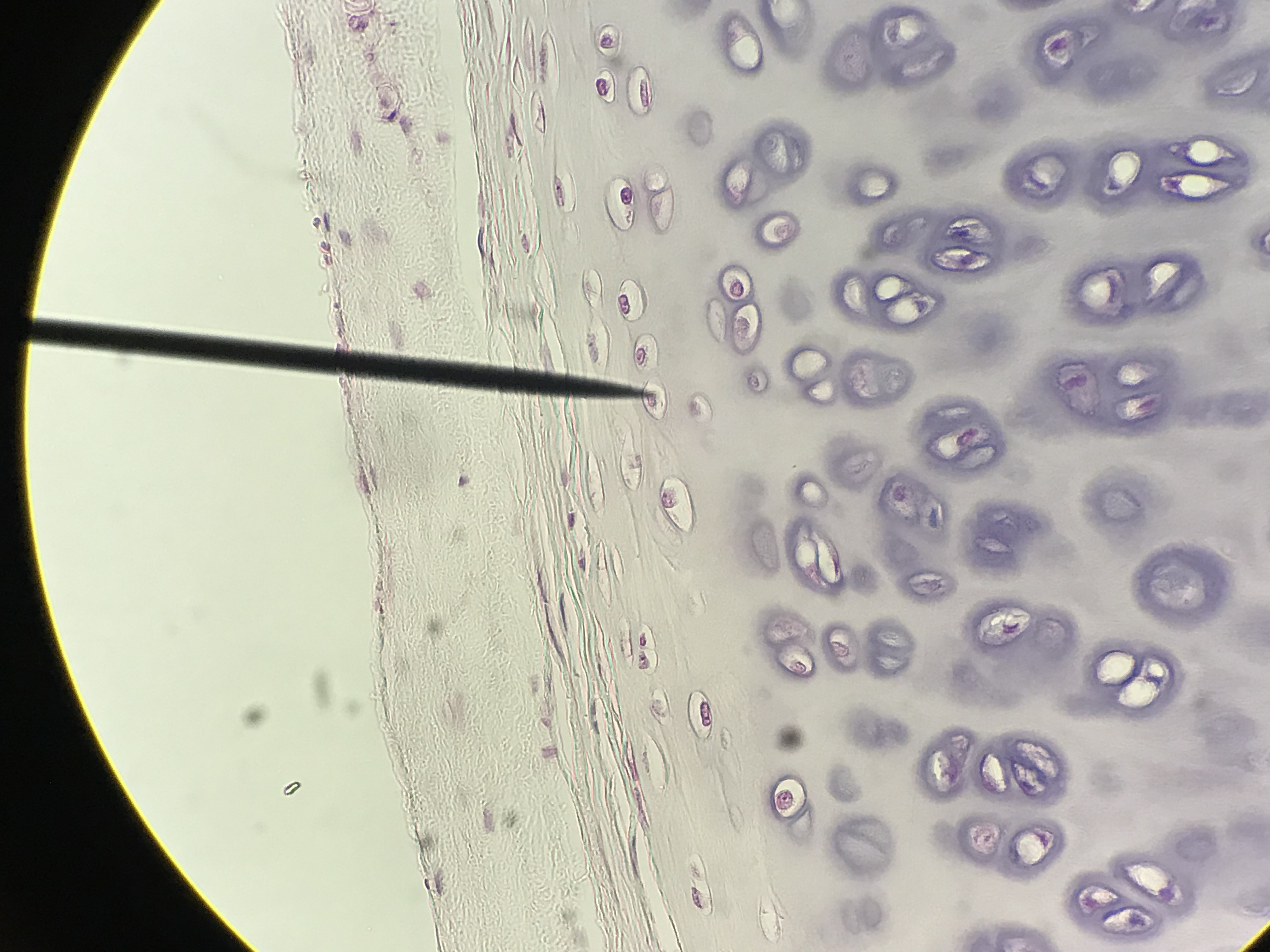
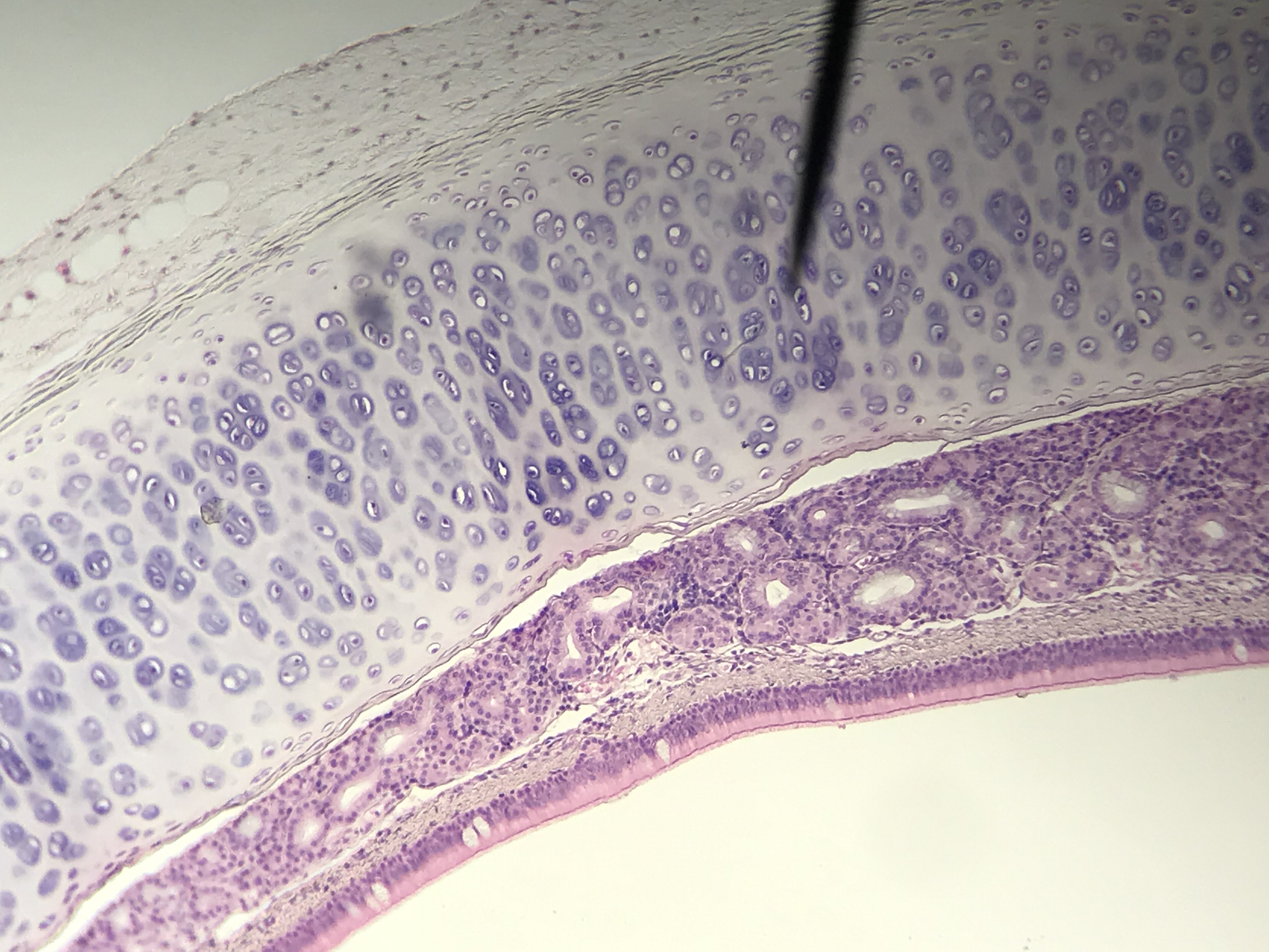
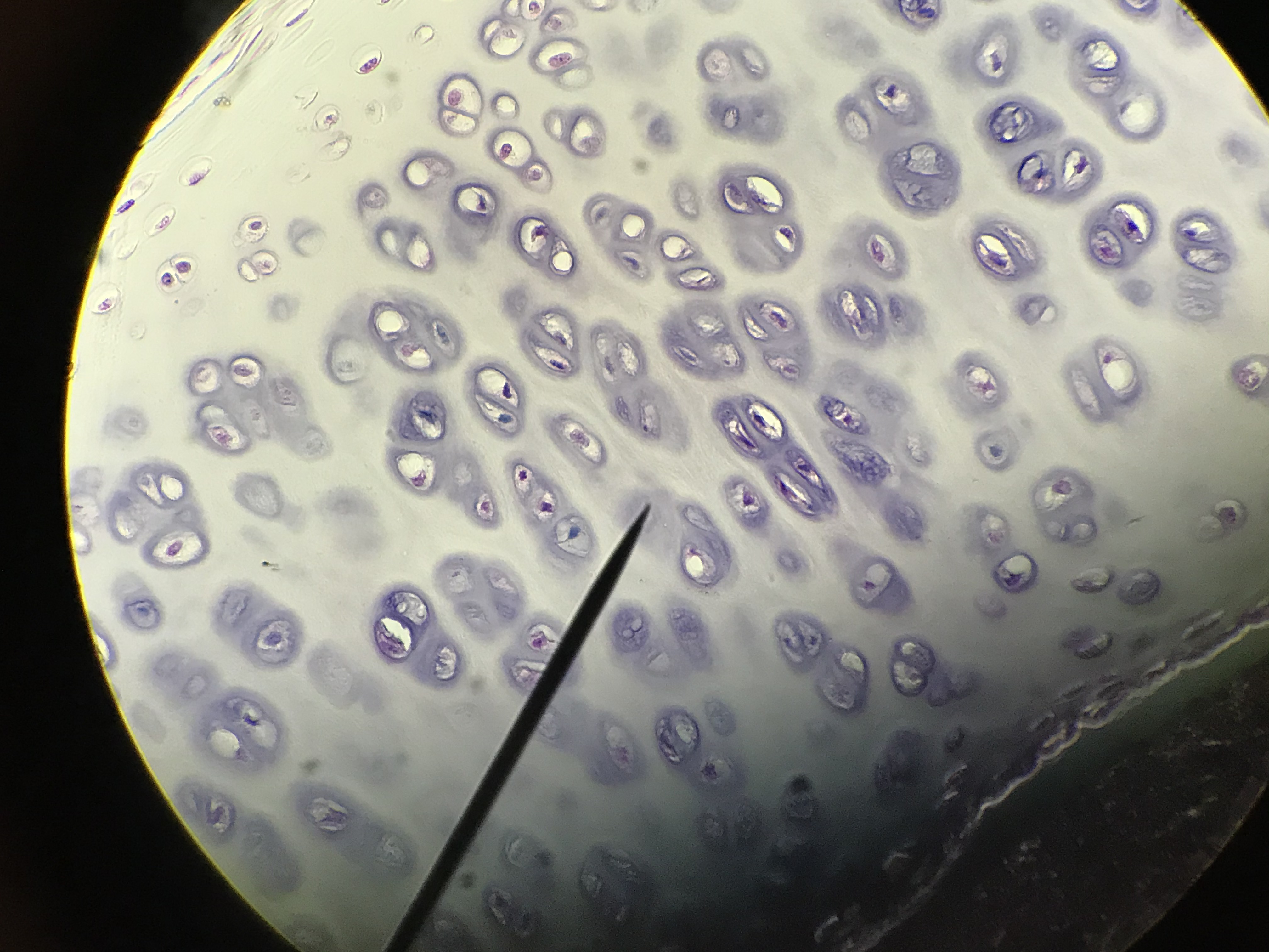
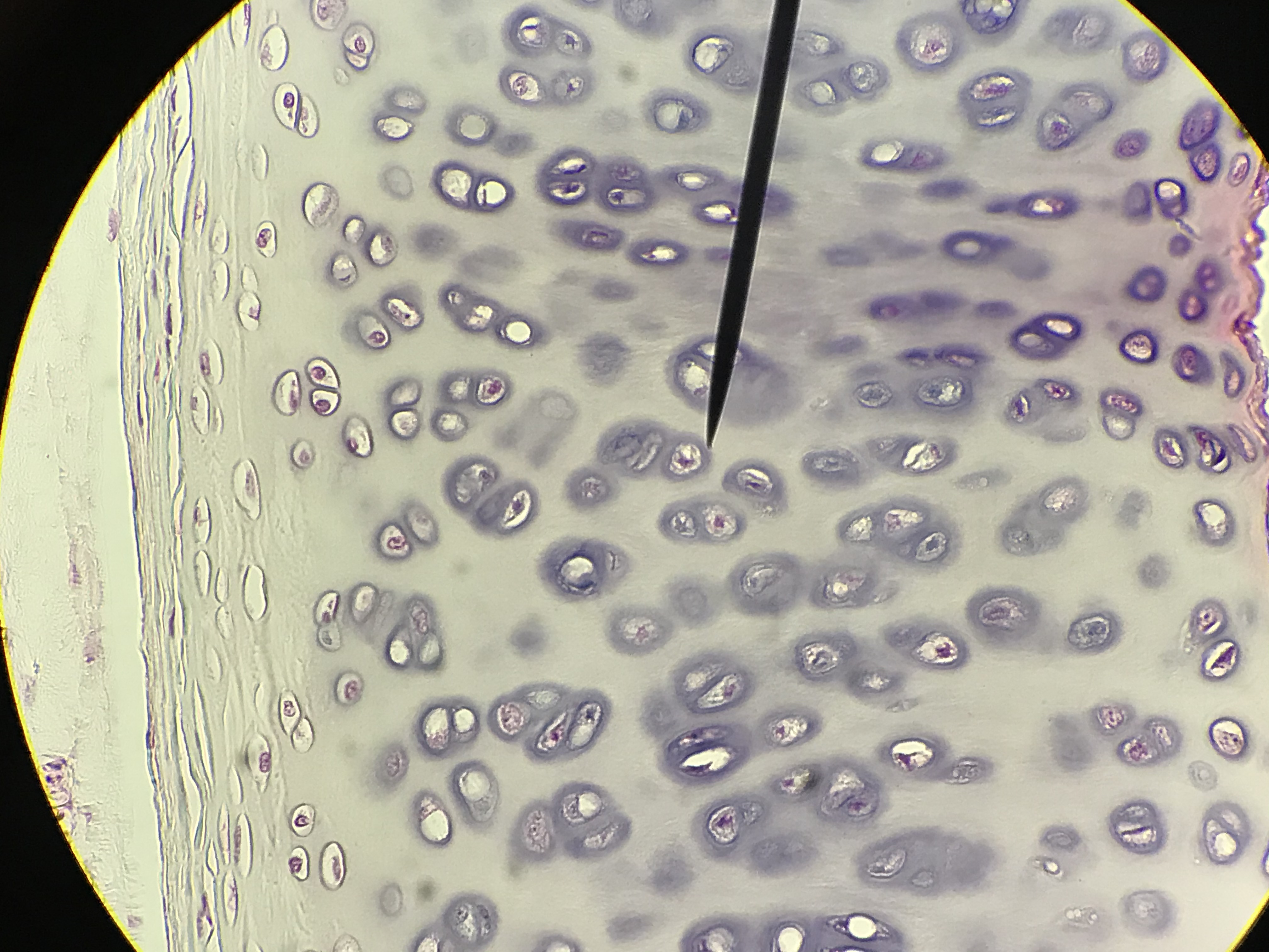
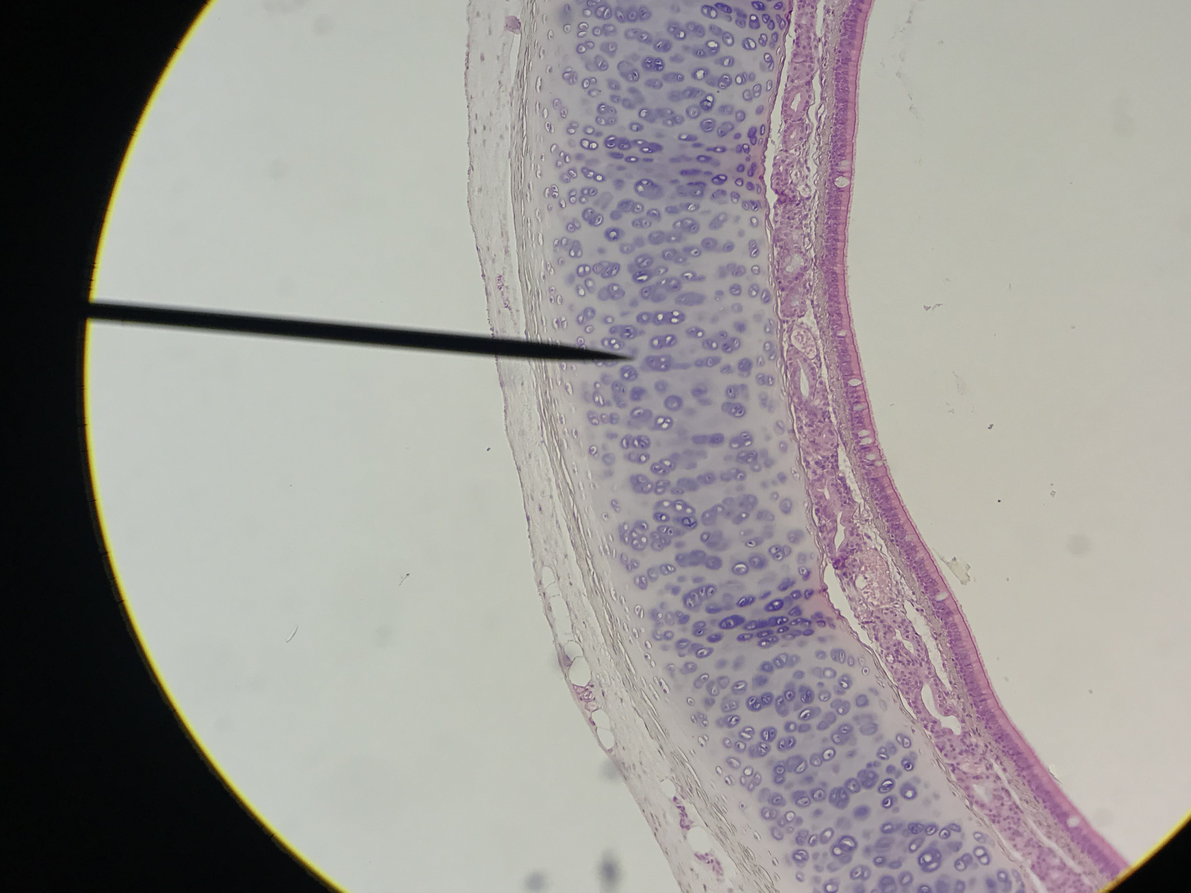
When observing all three types of cartilage under the microscope, the most dominate feature is the matrix which looks like a purple or blue "jelly". The matrix of all three types of cartilage has type II collagen fibers in it but these fibers are not visible with the stain we use. Inside of the matrix, cavities called lacuna can be observed. Mature cartilage cells, called chondrocytes, are found in the lacuna and they produce and maintain the matrix. Hyaline and elastic cartilage are both surrounded by a sheath of dense irregular connective tissue called the perichondrium. Within the fibers of the perichondrium are chondroblasts which are cartilage building cells. In adults, these cells are typically inactive but they can repair the cartilage if damaged. In children, chondroblasts allow for appositional growth (thickening) of the cartilage. Hyaline cartilage is the most abundant cartilage and it does not have any additional histological features. Most bone start out as hyaline
Slides on this page were made by students between the spring of 2018 and the spring of 2020. Go through
the diffrent student pictures and compare them to your lab book picture. Then slect one to draw on paper. Be sure
to label the cells, lacuna, fibers and other structures.
| Lab Book Image |
Student Images |
|
1 / 8
2 / 8
3/ 8
4 / 8
5 / 8
6 / 8
7 / 8
8 / 8
❮
❯
|