Panoramic Models
Head 1
Miniman 1
Miniman 2
Now identify on models
 Cephalic (head) regions |
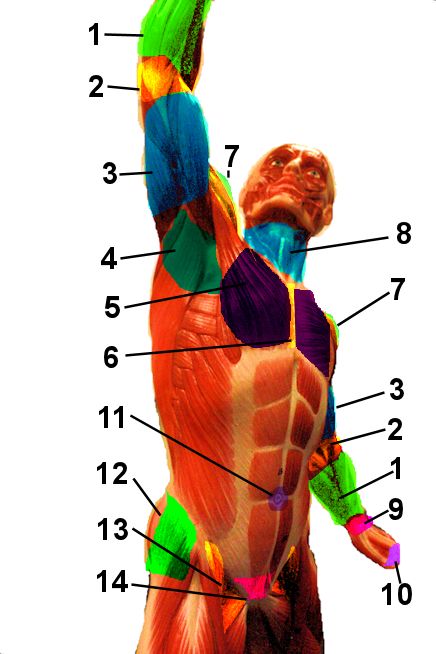
 Regions of the torso |
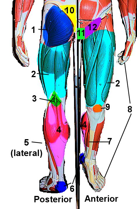
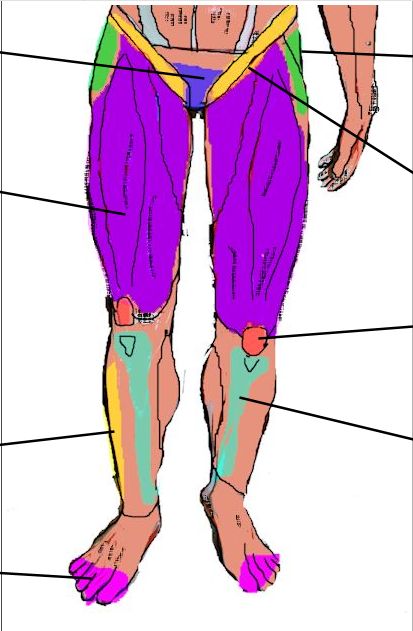
 Regions of the legs |
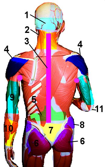
 Posterior Regions |
Sticky Notes
Head |
Torso |
Leg |
Back |
Panoramic Models |
||||||
|---|---|---|---|---|---|---|
| Rotate the models. Try to find all the anatomical regions talked about in your lab book on page 4. When you think you know the regions, try the sticky note activities. | ||||||
Head 1 |
Miniman 1 |
Miniman 2 |
Now identify on models |
|||
| Click on the links below. Try to identify the colored regions. then put your mouse over the region and see if you are right. | ||||||
|
||||||
Sticky Notes |
||||||
| Open each page. Place the sticky notes so they match to the correct region. Screen shot your answer (fn+ print screen on pc, command+shift+3 on mac) Upload your answers to Canvas | ||||||
| ||||||