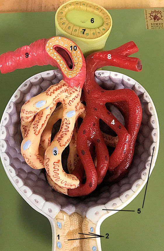
This is a model of the glomerulus. It is the 3rd part of the kidney series model showing the microscopic anatomy of
the glomerulus and associated structures. I like this one because you can really
see the difference between the afferent and efferent arterioloes and you can also see the podocytes. YOu can even see the microvilli on the proximal convoluted tubule.
Try to identify the structures given. You may want to compare this to the renal cortex histology. Be sure to see
the whole kidney and the nephron level from the kidney series model.