Word Bank
- Biceps Femoris
- Extensor Digitorum
- Fibularis Brevis
- Fibularis Longus
- Flexor Digitorum Longus
- Gastrocnemius
- Iliotibial Band/Tract
- Sartorius
- Semimembranosus
- Semitendinosus
- Soleus
- Tibialis Anterior
- Vastus lateralis
- Vastus Medialis
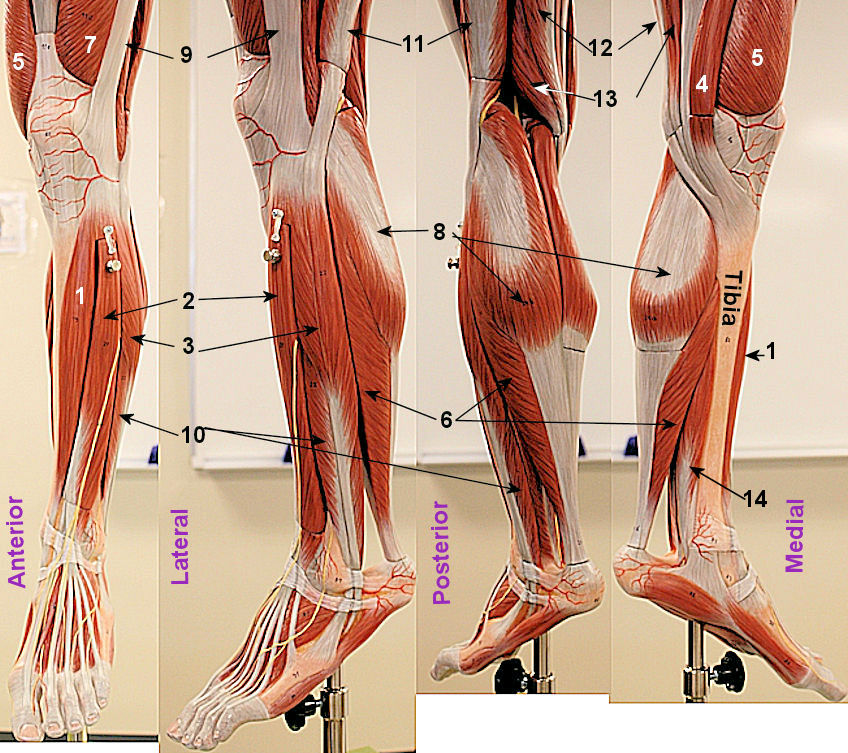
The next lab exam will be identifying muscles. I will give you a model and ask you what muscle I labeled on it. The pictures on this page are of the left calf from 4 viws; anterior, lateral, posterior, and medial. I tried
to use the same number for the same muscles. As a result some numbers are between models pointing to two models. When done, try the hip flexors, the deep posterior hip are seperate pages as are the
thigh muscles.