Lab 4 Skin models |
||
|---|---|---|
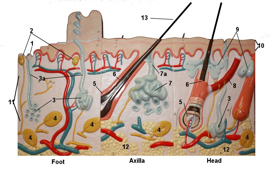 Skin Model with Burns on Back |
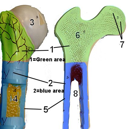
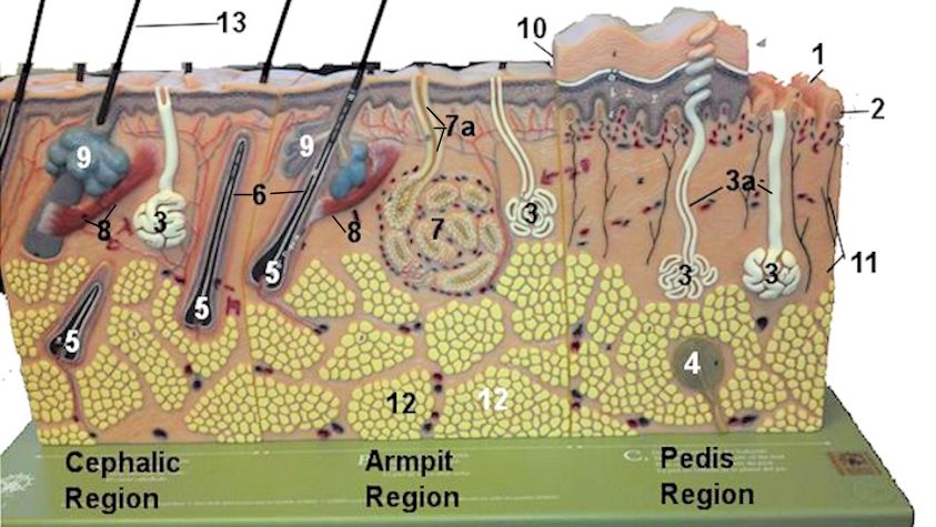 Skin model on Green Base |
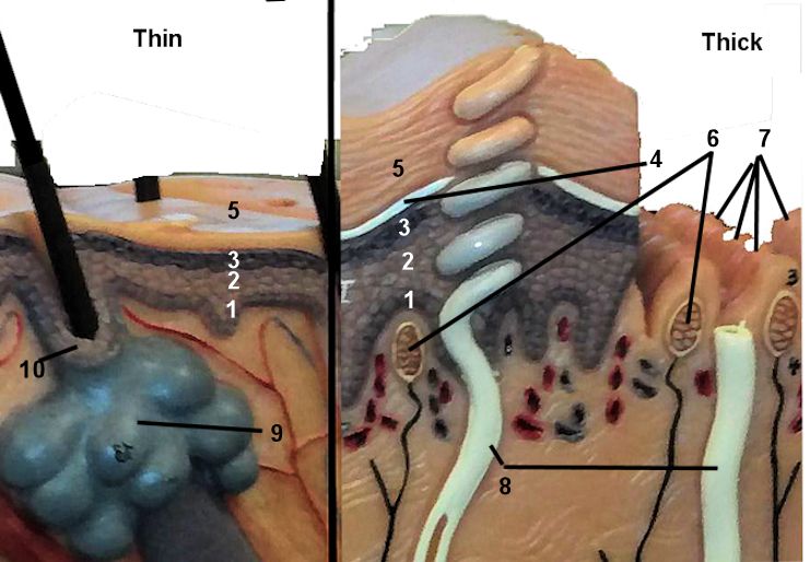 Epidermis on model |
Lab 4 Epidermis Histology |
||
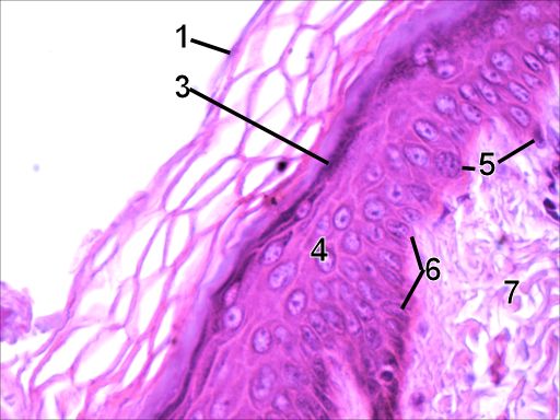
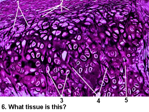
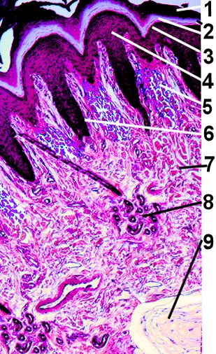 Epidermis of Thick Skin |
 Epidermis of Thin Skin |
Please see the study guide on Canvas for more histology. I will ask you to identify specific structures of the dermis. |
Lab 5 Bone models |
||
 Longbone Model |
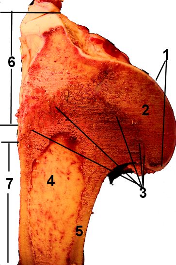 Cow Humerus |
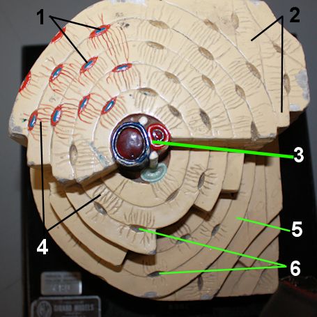 Osteon Model |
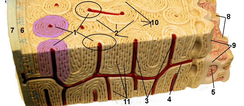 Compact Bone Model |
Lab 5 Supporting Connective Tissue Histology |
|
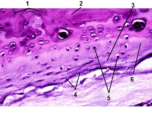
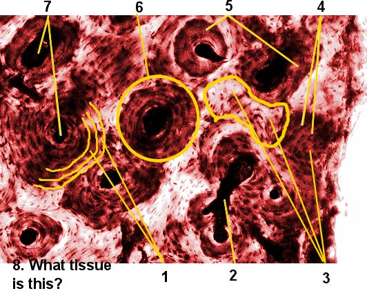 Compact bone |
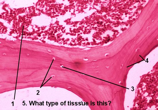 Spongy bone |
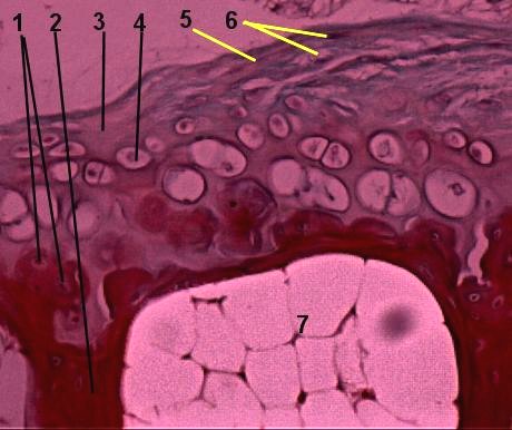
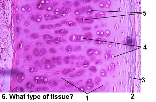 Hyaline Cartilage |
 Elastic Cartilage |
 Fibrocartilage easy |
 Fibrocartilage hard but with proper colors |
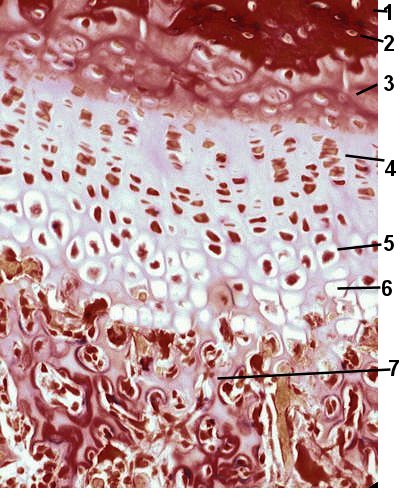 Zones of Growth Plate |
||